

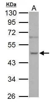
Sample (30 μg of whole cell lysate) A: PC-3 10% SDS PAGE GTX102831 diluted at 1:1000 The HRP-conjugated anti-rabbit IgG antibody (GTX213110-01) was used to detect the primary antibody.
![NOVA1 antibody detects NOVA1 protein by immunofluorescent analysis. Sample: DIV9 rat E18 primary hippocampal neuron cells were fixed in 4% paraformaldehyde at RT for 15 min. Green: NOVA1 stained by NOVA1 antibody (GTX102831) diluted at 1:500. Red: beta Tubulin 3/ Tuj1, stained by beta Tubulin 3/ Tuj1 antibody [GT11710] (GTX631836) diluted at 1:500. Blue: Fluoroshield with DAPI (GTX30920).](https://www.genetex.com/upload/website/prouct_img/normal/GTX102831/GTX102831_39974_20181004_ICC_IF_R_w_23060119_449.webp "NOVA1 antibody detects NOVA1 protein by immunofluorescent analysis. Sample: DIV9 rat E18 primary hippocampal neuron cells were fixed in 4% paraformaldehyde at RT for 15 min. Green: NOVA1 stained by NOVA1 antibody (GTX102831) diluted at 1:500. Red: beta Tubulin 3/ Tuj1, stained by beta Tubulin 3/ Tuj1 antibody [GT11710] (GTX631836) diluted at 1:500. Blue: Fluoroshield with DAPI (GTX30920).")
Sample (30 μg of whole cell lysate) A: PC-3 10% SDS PAGE GTX102831 diluted at 1:1000 The HRP-conjugated anti-rabbit IgG antibody (GTX213110-01) was used to detect the primary antibody.
NOVA1 antibody
GTX102831
ApplicationsImmunoFluorescence, Western Blot, ImmunoCytoChemistry
Product group Antibodies
ReactivityHuman
TargetNOVA1
Overview
- SupplierGeneTex
- Product NameNOVA1 antibody
- Delivery Days Customer9
- Application Supplier NoteWB: 1:500-1:3000. ICC/IF: 1:100-1:1000. *Optimal dilutions/concentrations should be determined by the researcher.Not tested in other applications.
- ApplicationsImmunoFluorescence, Western Blot, ImmunoCytoChemistry
- CertificationResearch Use Only
- ClonalityPolyclonal
- Concentration0.92 mg/ml
- ConjugateUnconjugated
- Gene ID4857
- Target nameNOVA1
- Target descriptionNOVA alternative splicing regulator 1
- Target synonymsNova-1, RNA-binding protein Nova-1, neuro-oncological ventral antigen 1, onconeural ventral antigen 1, paraneoplastic Ri antigen, ventral neuron-specific protein 1
- HostRabbit
- IsotypeIgG
- Protein IDP51513
- Protein NameRNA-binding protein Nova-1
- Scientific DescriptionThis gene encodes a neuron-specific RNA-binding protein, a member of the Nova family of paraneoplastic disease antigens, that is recognized and inhibited by paraneoplastic antibodies. These antibodies are found in the sera of patients with paraneoplastic opsoclonus-ataxia, breast cancer, and small cell lung cancer. Alternatively spliced transcripts encoding distinct isoforms have been described. [provided by RefSeq]
- ReactivityHuman
- Storage Instruction-20°C or -80°C,2°C to 8°C
- UNSPSC41116161
Datasheet
Related products
Product group Antibodies
NOVA1 AntibodyCSB-PA015957LA01HU
ApplicationsWestern Blot, ELISA, ImmunoHistoChemistry
ReactivityHuman
TargetNOVA1
- SizePrice

Product group Antibodies
Anti-Nova1 Antibody Picoband(r)A06087-1-CARRIER-FREE
ApplicationsFlow Cytometry, ImmunoFluorescence, Western Blot, ELISA, ImmunoCytoChemistry, ImmunoHistoChemistry, ImmunoHistoChemistry Frozen
ReactivityHuman, Mouse, Rat
TargetNOVA1
- SizePrice
Product group Antibodies

NOVA1 AntibodyLS-C747238
ApplicationsWestern Blot
ReactivityHuman
TargetNOVA1
- SizePrice
Product group Antibodies

Goat anti-NOVA1EB08387
ApplicationsWestern Blot, ELISA, ImmunoHistoChemistry
ReactivityHuman
TargetNOVA1
- SizePrice

Product group Antibodies
Anti-NOVA1 AntibodyHPA004155
ApplicationsWestern Blot, ImmunoCytoChemistry, ImmunoHistoChemistry
ReactivityHuman
TargetNOVA1
- SizePrice
Product group Antibodies
NOVA1 Polyclonal AntibodyCAC13137
ApplicationsWestern Blot, ELISA, ImmunoHistoChemistry
TargetNOVA1
- SizePrice
Product group Antibodies
NOVA1 antibody, InternalGTX88815
ApplicationsWestern Blot, ImmunoHistoChemistry, ImmunoHistoChemistry Paraffin
ReactivityHuman
TargetNOVA1
- SizePrice
Product group Antibodies
Anti-NOVA1 (Center) Antibody102-24148
ApplicationsWestern Blot, ImmunoHistoChemistry, ImmunoHistoChemistry Paraffin
TargetNOVA1
- SizePrice