

![p27 / KIP1(SX53G8), Biotin conjugate, 0.1mg/mL [26628-22-8]](https://biotium.com/wp-content/uploads/2019/02/P27-SX53G8-CF488A-GAM-Phalloidin-CF633-PFA-MCF7-011619.jpg "p27 / KIP1(SX53G8), Biotin conjugate, 0.1mg/mL [26628-22-8]")
![p27 / KIP1(SX53G8), Biotin conjugate, 0.1mg/mL [26628-22-8]](https://biotium.com/wp-content/uploads/2016/12/BNUB0669-1-1.jpg "p27 / KIP1(SX53G8), Biotin conjugate, 0.1mg/mL [26628-22-8]")
![p27 / KIP1(SX53G8), Biotin conjugate, 0.1mg/mL [26628-22-8]](https://biotium.com/wp-content/uploads/2016/12/BNUB0669-2-1-e1612211382555.jpg "p27 / KIP1(SX53G8), Biotin conjugate, 0.1mg/mL [26628-22-8]")
p27 / KIP1(SX53G8), Biotin conjugate, 0.1mg/mL [26628-22-8]
BNCB0669
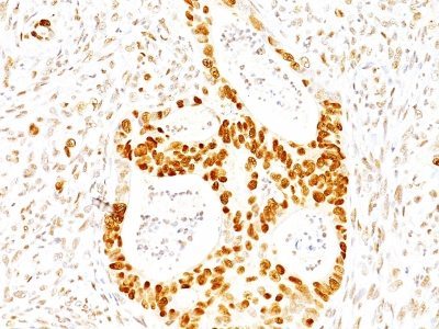
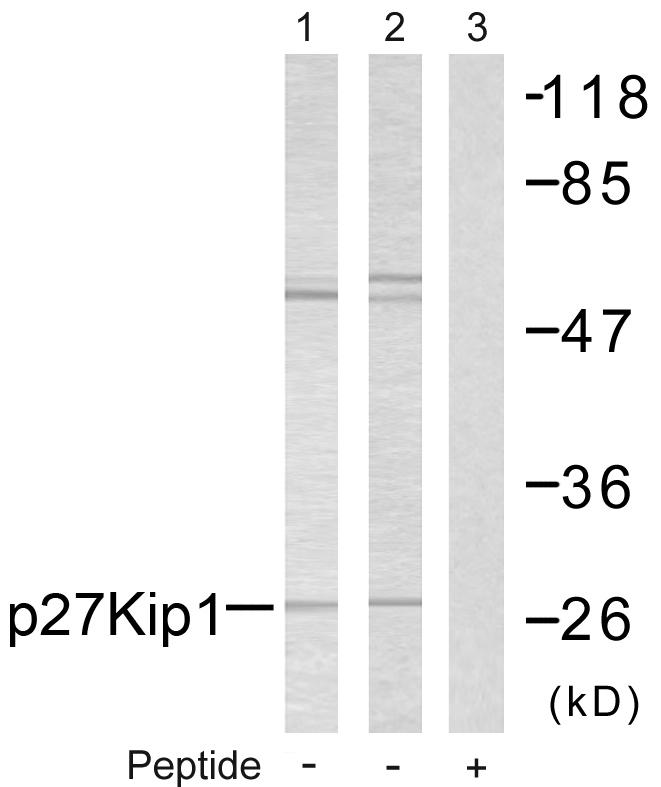
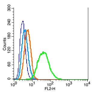
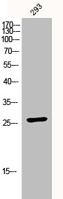
ApplicationsFlow Cytometry, ImmunoFluorescence, ImmunoPrecipitation, Western Blot, ImmunoHistoChemistry, ImmunoHistoChemistry Paraffin
Product group Antibodies
ReactivityBovine, Human, Monkey, Mouse, Rat
TargetCDKN1B
Overview
- SupplierBiotium
- Product Namep27 / KIP1(SX53G8), Biotin conjugate, 0.1mg/mL [26628-22-8]
- Delivery Days Customer9
- ApplicationsFlow Cytometry, ImmunoFluorescence, ImmunoPrecipitation, Western Blot, ImmunoHistoChemistry, ImmunoHistoChemistry Paraffin
- CAS Number26628-22-8
- CertificationResearch Use Only
- ClonalityMonoclonal
- Clone IDSX53G8
- Concentration0.1 mg/ml
- ConjugateBiotin
- Gene ID1027
- Target nameCDKN1B
- Target descriptioncyclin dependent kinase inhibitor 1B
- Target synonymsCDKN4, KIP1, MEN1B, MEN4, P27KIP1, cyclin-dependent kinase inhibitor 1B, cyclin-dependent kinase inhibitor 1B (p27, Kip1)
- HostMouse
- IsotypeIgG1
- Protein IDP46527
- Protein NameCyclin-dependent kinase inhibitor 1B
- Scientific DescriptionThis MAb recognizes a 27 kDa protein, identified as the p27Kip1, a cell cycle regulatory mitotic inhibitor. It is highly specific and shows no cross-reaction with other related mitotic inhibitors. In Western blotting of cell lysates from 7 human breast cancer cell lines (ZR75-1, ZR75-30, MCF-7, MDAMB453, T47D, CAL51, 734B), the antibody labels a single band corresponding to p27Kip1. It functions as a negative regulator of G1 progression and has been proposed to function as a possible mediator of TGF-betanduced G1 arrest. p27Kip1 is a candidate tumor suppressor gene. Reportedly, low p27 expression has been associated with unfavorable prognosis in renal cell carcinoma, colon carcinoma, breast carcinomas, non-small-cell lung carcinoma, hepatocellular carcinoma, multiple myeloma, and lymph node metastases in papillary carcinoma of the thyroid, as well as a more aggressive phenotype in carcinoma of the cervix.Primary antibodies are available purified, or with a selection of fluorescent CF® Dyes and other labels. CF® Dyes offer exceptional brightness and photostability. Note: Conjugates of blue fluorescent dyes like CF®405S and CF®405M are not recommended for detecting low abundance targets, because blue dyes have lower fluorescence and can give higher non-specific background than other dye colors.
- SourceAnimal
- ReactivityBovine, Human, Monkey, Mouse, Rat
- Storage Instruction2°C to 8°C,RT
- UNSPSC41116161
MSDS
Related products
Product group Antibodies
Anti-p27 Kip1 AntibodyA95332
ApplicationsWestern Blot, ELISA, ImmunoHistoChemistry
ReactivityHuman, Mouse, Rat
- SizePrice
Product group Antibodies
Anti-p27 Antibody130-10050
ApplicationsELISA
ReactivityHuman
TargetCDKN1B
- SizePrice
Product group Antibodies
References
ApplicationsFlow Cytometry, ImmunoFluorescence, Western Blot, ELISA, ImmunoCytoChemistry, ImmunoHistoChemistry, ImmunoHistoChemistry Frozen, ImmunoHistoChemistry Paraffin
ReactivityCanine, Chicken, Human, Mouse, Porcine, Rat, Sheep
TargetCDKN1B
- SizePrice
Product group Antibodies
CDKN1B AntibodyCSB-PA003625
ApplicationsWestern Blot, ELISA, ImmunoHistoChemistry
ReactivityHuman, Mouse, Rat
TargetCDKN1B
- SizePrice
Product group Antibodies

ApplicationsImmunoPrecipitation, Western Blot, ELISA
ReactivityBovine, Canine, Human, Mouse, Rat
TargetCDKN1B
- SizePrice
Product group Antibodies
ApplicationsWestern Blot, ImmunoHistoChemistry
ReactivityMouse, Rat
TargetCDKN1B
- SizePrice
Product group Antibodies

ApplicationsELISA
ReactivityHuman
TargetCDKN1B
- SizePrice
Product group Antibodies
p27 Kip1 antibodyGTX100446
ApplicationsImmunoFluorescence, ImmunoPrecipitation, Western Blot, ImmunoCytoChemistry, ImmunoHistoChemistry, ImmunoHistoChemistry Frozen, ImmunoHistoChemistry Paraffin
ReactivityHuman, Mouse, Rat
TargetCDKN1B
- SizePrice

Product group Antibodies
Anti-CDKN1B AntibodyHPA059086
ApplicationsWestern Blot, ImmunoCytoChemistry
ReactivityHuman
TargetCDKN1B
- SizePrice