

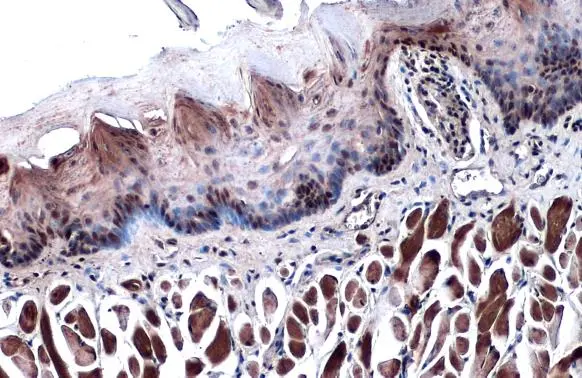
p63 antibody [N2C1], Internal detects p63 protein at nucleus by immunohistochemical analysis. Sample: Paraffin-embedded mouse tongue. p63 stained by p63 antibody [N2C1], Internal (GTX102425) diluted at 1:500. Antigen Retrieval: Citrate buffer, pH 6.0, 15 min
![p63 antibody [N2C1], Internal detects TP63 protein by western blot analysis. A. 50 μg rat brain lysate/extract 7.5% SDS-PAGE p63 antibody [N2C1], Internal (GTX102425) dilution: 1:500 The HRP-conjugated anti-rabbit IgG antibody (GTX213110-01) was used to detect the primary antibody.](https://www.genetex.com/upload/website/prouct_img/normal/GTX102425/GTX102425_39918_WB_R_brain_w_23060100_846.webp "p63 antibody [N2C1], Internal detects TP63 protein by western blot analysis. A. 50 μg rat brain lysate/extract 7.5% SDS-PAGE p63 antibody [N2C1], Internal (GTX102425) dilution: 1:500 The HRP-conjugated anti-rabbit IgG antibody (GTX213110-01) was used to detect the primary antibody.")
, p63 (GTX102425) and Cytokeratin 7 (GTX109723) in human small cell lung cancer (SCLC) and non-small cell lung cancer (NSCLC) specimens. Sample: Paraffin-embedded human SCLC (upper panel) and NSCLC (lower panel). The section was pre-treated using heat mediated antigen retrieval with sodium citrate buffer (pH6) for 15 mins. The section was then incubated with primary antibody at 1:500 overnight at 4oC and detected using an HRP conjugated avidin-biotin-peroxidase Complex system. DAB was used as the chromogen and counterstained with haematoxylin.
Antigen Retrieval: Citrate buffer, pH 6.0, 15 min")
 antibody at 1:100 dilution.
Antigen Retrieval: Trilogy? (EDTA based, pH 8.0) buffer, 15min")
![Non-transfected (–) and transfected (+) A431 whole cell extracts (30 μg) were separated by 7.5% SDS-PAGE, and the membrane was blotted with p63 antibody [N2C1], Internal (GTX102425) diluted at 1:2000. The HRP-conjugated anti-rabbit IgG antibody (GTX213110-01) was used to detect the primary antibody.](https://www.genetex.com/upload/website/prouct_img/normal/GTX102425/GTX102425_41472_20210716_WB_shRNA_watermark_w_23060100_893.webp "Non-transfected (–) and transfected (+) A431 whole cell extracts (30 μg) were separated by 7.5% SDS-PAGE, and the membrane was blotted with p63 antibody [N2C1], Internal (GTX102425) diluted at 1:2000. The HRP-conjugated anti-rabbit IgG antibody (GTX213110-01) was used to detect the primary antibody.")
![p63 antibody [N2C1], Internal detects TP63 protein by western blot analysis. A. 50 μg mouse brain lysate/extract 7.5% SDS-PAGE p63 antibody [N2C1], Internal (GTX102425) dilution: 1:500 The HRP-conjugated anti-rabbit IgG antibody (GTX213110-01) was used to detect the primary antibody.](https://www.genetex.com/upload/website/prouct_img/normal/GTX102425/GTX102425_39918_WB_M_brain_w_23060100_558.webp "p63 antibody [N2C1], Internal detects TP63 protein by western blot analysis. A. 50 μg mouse brain lysate/extract 7.5% SDS-PAGE p63 antibody [N2C1], Internal (GTX102425) dilution: 1:500 The HRP-conjugated anti-rabbit IgG antibody (GTX213110-01) was used to detect the primary antibody.")
![Immunoprecipitation of p63 protein from A431 whole cell extracts using 5 μg of p63 antibody [N2C1], Internal (GTX102425). Western blot analysis was performed using p63 antibody [N2C1], Internal (GTX102425). EasyBlot anti-Rabbit IgG (GTX221666-01) was used as a secondary reagent.](https://www.genetex.com/upload/website/prouct_img/normal/GTX102425/GTX102425_39918_20150910_IP_w_23060100_766.webp "Immunoprecipitation of p63 protein from A431 whole cell extracts using 5 μg of p63 antibody [N2C1], Internal (GTX102425). Western blot analysis was performed using p63 antibody [N2C1], Internal (GTX102425). EasyBlot anti-Rabbit IgG (GTX221666-01) was used as a secondary reagent.")
![p63 antibody [N2C1], Internal detects p63 protein at nucleus by immunofluorescent analysis. Sample: A431 cells were fixed in 4% paraformaldehyde at RT for 15 min. Green: p63 stained by p63 antibody [N2C1], Internal (GTX102425) diluted at 1:500. Red: alpha Tubulin, stained by alpha Tubulin antibody [GT114] (GTX628802) diluted at 1:500.](https://www.genetex.com/upload/website/prouct_img/normal/GTX102425/GTX102425_43138_20190327_ICC_IF_w_23060100_737.webp "p63 antibody [N2C1], Internal detects p63 protein at nucleus by immunofluorescent analysis. Sample: A431 cells were fixed in 4% paraformaldehyde at RT for 15 min. Green: p63 stained by p63 antibody [N2C1], Internal (GTX102425) diluted at 1:500. Red: alpha Tubulin, stained by alpha Tubulin antibody [GT114] (GTX628802) diluted at 1:500.")


p63 antibody [N2C1], Internal detects p63 protein at nucleus by immunohistochemical analysis. Sample: Paraffin-embedded mouse tongue. p63 stained by p63 antibody [N2C1], Internal (GTX102425) diluted at 1:500. Antigen Retrieval: Citrate buffer, pH 6.0, 15 min
p63 antibody [N2C1], Internal
GTX102425
ApplicationsImmunoFluorescence, ImmunoPrecipitation, Western Blot, ImmunoCytoChemistry, ImmunoHistoChemistry, ImmunoHistoChemistry Frozen, ImmunoHistoChemistry Paraffin, Other Application
Product group Antibodies
ReactivityCanine, Feline, Human, Mouse, Rat
TargetTP63
Overview
- SupplierGeneTex
- Product Namep63 antibody [N2C1], Internal
- Delivery Days Customer9
- Application Supplier NoteWB: 1:500-1:3000. ICC/IF: 1:100-1:1000. IHC-P: 1:100-1:1000. IP: 1:100-1:500. *Optimal dilutions/concentrations should be determined by the researcher.Not tested in other applications.
- ApplicationsImmunoFluorescence, ImmunoPrecipitation, Western Blot, ImmunoCytoChemistry, ImmunoHistoChemistry, ImmunoHistoChemistry Frozen, ImmunoHistoChemistry Paraffin, Other Application
- CertificationResearch Use Only
- ClonalityPolyclonal
- Concentration1.37 mg/ml
- ConjugateUnconjugated
- Gene ID8626
- Target nameTP63
- Target descriptiontumor protein p63
- Target synonymsAIS, B(p51A), B(p51B), EEC3, KET, LMS, NBP, OFC8, RHS, SHFM4, TP53CP, TP53L, TP73L, p40, p51, p53CP, p63, p73H, p73L, tumor protein 63, amplified in squamous cell carcinoma, chronic ulcerative stomatitis protein, keratinocyte transcription factor KET, transformation-related protein 63, tumor protein p53-competing protein
- HostRabbit
- IsotypeIgG
- Protein IDQ9H3D4
- Protein NameTumor protein 63
- Scientific DescriptionThis gene encodes a member of the p53 family of transcription factors. An animal model, p63 -/- mice, has been useful in defining the role this protein plays in the development and maintenance of stratified epithelial tissues. p63 -/- mice have several developmental defects which include the lack of limbs and other tissues, such as teeth and mammary glands, which develop as a result of interactions between mesenchyme and epithelium. Mutations in this gene are associated with ectodermal dysplasia, and cleft lip/palate syndrome 3 (EEC3); split-hand/foot malformation 4 (SHFM4); ankyloblepharon-ectodermal defects-cleft lip/palate; ADULT syndrome (acro-dermato-ungual-lacrimal-tooth); limb-mammary syndrome; Rap-Hodgkin syndrome (RHS); and orofacial cleft 8. Both alternative splicing and the use of alternative promoters results in multiple transcript variants encoding different proteins. Many transcripts encoding different proteins have been reported but the biological validity and the full-length nature of these variants have not been determined. [provided by RefSeq]
- ReactivityCanine, Feline, Human, Mouse, Rat
- Storage Instruction-20°C or -80°C,2°C to 8°C
- UNSPSC12352203
References
- Yamamoto S, Matsui K, Kinoshita Y, et al. Successful reconstruction of the rat ureter by a syngeneic collagen tube with a cardiomyocyte sheet. Regen Ther. 2023,24:561-567. doi: 10.1016/j.reth.2023.10.001Read this paper
- Lee JM, Lim TY, Oh SB, et al. Ahnak is required to balance calcium ion homeostasis and smooth muscle development in the urinary system. Cell Biosci. 2023,13(1):108. doi: 10.1186/s13578-023-01055-xRead this paper
- Russell SK, Harrison JK, Olson BS, et al. Uropathogenic Escherichia coli infection-induced epithelial trained immunity impacts urinary tract disease outcome. Nat Microbiol. 2023,8(5):875-888. doi: 10.1038/s41564-023-01346-6Read this paper
- Neumayer G, Torkelson JL, Li S, et al. A scalable, GMP-compatible, autologous organotypic cell therapy for Dystrophic Epidermolysis Bullosa. bioRxiv. 2023,:pii: 2023.02.28.529447. doi: 10.1101/2023.02.28.529447.Read this paper
- Bedos L, Wickham H, Gabriel V, et al. Culture and characterization of canine and feline corneal epithelial organoids: A new tool for the study and treatment of corneal diseases. Front Vet Sci. 2022,9:1050467. doi: 10.3389/fvets.2022.1050467Read this paper
- Huang H, Fang Y, Jiang M, et al. Contribution of Trp63(CreERT2)-labeled cells to alveolar regeneration is independent of tuft cells. Elife. 2022,11. doi: 10.7554/eLife.78217Read this paper
- Huang H, Liu J, Li M, et al. Cis-regulatory chromatin loops analysis identifies GRHL3 as a master regulator of surface epithelium commitment. Sci Adv. 2022,8(28):eabo5668. doi: 10.1126/sciadv.abo5668Read this paper
- Kadur Lakshminarasimha Murthy P, Sontake V, Tata A, et al. Human distal lung maps and lineage hierarchies reveal a bipotent progenitor. Nature. 2022,604(7904):111-119. doi: 10.1038/s41586-022-04541-3Read this paper
- Chalmers FE, Dusold JE, Shaik JA, et al. Targeted deletion of TGFβ1 in basal keratinocytes causes profound defects in stratified squamous epithelia and aberrant melanocyte migration. Dev Biol. 2022,485:9-23. doi: 10.1016/j.ydbio.2022.02.009Read this paper
- Mou H, Yang Y, Riehs MA, et al. Airway basal stem cells generate distinct subpopulations of PNECs. Cell Rep. 2021,35(3):109011. doi: 10.1016/j.celrep.2021.109011Read this paper
Datasheet
Related products
Product group Antibodies

Anti-p63 [BU5]Ab00871-10.0
ApplicationsFlow Cytometry, ImmunoFluorescence
ReactivityHuman
TargetTP63
- SizePrice

Product group Antibodies
Anti-TP63 Antibody144-12937
ApplicationsImmunoFluorescence, Western Blot
ReactivityHuman, Mouse, Rat
TargetTP63
- SizePrice
![ICC/IF analysis of PFA-fixed HeLa cells using GTX02728 p63 antibody [TP40/3980R]. Green : Primary antibody Red : Phalloiden (membrane)](https://www.genetex.com/upload/website/prouct_img/normal/GTX02728/GTX02728_20210319_ICCIF_w_23053122_851.webp)
Product group Antibodies
p63 antibody [TP40/3980R]GTX02728
ApplicationsFlow Cytometry, ImmunoFluorescence, ImmunoCytoChemistry, ImmunoHistoChemistry, ImmunoHistoChemistry Paraffin
ReactivityHuman
TargetTP63
- SizePrice
Product group Antibodies
p63 antibody [DBR16.1]GTX03308
ApplicationsImmunoHistoChemistry, ImmunoHistoChemistry Paraffin
ReactivityHuman
TargetTP63
- SizePrice
![IHC-P analysis of human esophagus tissue using GTX04458 p63 antibody [MSVA-063R] HistoMAX?. A strong p63 staining occurs squamous epithelial cells.](https://www.genetex.com/upload/website/prouct_img/normal/GTX04458/GTX04458_20230728_IHC-P_91_23072722_588.webp)
Product group Antibodies
ApplicationsImmunoHistoChemistry, ImmunoHistoChemistry Paraffin
ReactivityHuman
TargetTP63
- SizePrice
![WB analysis of A431 (1), HeLa (2), Jurkat (3), THP-1 (4), NIH3T3 (5), Cos7 (6) and PC-12 (7) cell lysate using GTX60407 p63 antibody [4E5].](https://www.genetex.com/upload/website/prouct_img/normal/GTX60407/GTX60407_20170912_WB_w_23061123_286.webp)
Product group Antibodies
p63 antibody [4E5]GTX60407
ApplicationsWestern Blot, ELISA, ImmunoHistoChemistry, ImmunoHistoChemistry Paraffin
ReactivityHuman, Monkey, Mouse, Rat
TargetTP63
- SizePrice
![Mouse tissue extract (50 μg) was separated by 7.5% SDS-PAGE, and the membrane was blotted with p63 antibody [GT1179] (GTX633818) diluted at 1:2000. The HRP-conjugated anti-mouse IgG antibody (GTX213111-01) was used to detect the primary antibody.](https://www.genetex.com/upload/website/prouct_img/normal/GTX633818/GTX633818_42688_20220401_WB_M_skin_w_23061202_100.webp)
Product group Antibodies
References
p63 antibody [GT1179]GTX633818
ApplicationsImmunoFluorescence, Western Blot, ImmunoCytoChemistry, ImmunoHistoChemistry, ImmunoHistoChemistry Paraffin
ReactivityHuman, Mouse
TargetTP63
- SizePrice
![Non-transfected (–) and transfected (+) A431 whole cell extracts (30 μg) were separated by 7.5% SDS-PAGE, and the membrane was blotted with p63 antibody [HL1027] (GTX635841) diluted at 1:50000. The HRP-conjugated anti-rabbit IgG antibody (GTX213110-01) was used to detect the primary antibody, and the signal was developed with Trident ECL plus-Enhanced.](https://www.genetex.com/upload/website/prouct_img/normal/GTX635841/GTX635841_44340_20221104_WB_shRNA_watermark_22110919_252.webp)
Product group Antibodies
p63 antibody [HL1027]GTX635841
ApplicationsImmunoFluorescence, Western Blot, ImmunoCytoChemistry, ImmunoHistoChemistry, ImmunoHistoChemistry Paraffin
ReactivityHuman, Mouse, Zebra Fish
TargetTP63
- SizePrice