

PathPlus(tm) EPCAM Antibody (clone MOC-31, Concentrated)
LS-B5565
ApplicationsWestern Blot, ImmunoHistoChemistry, ImmunoHistoChemistry Frozen, ImmunoHistoChemistry Paraffin
Product group Antibodies
ReactivityHuman
TargetEPCAM
Overview
- SupplierLifeSpan BioSciences
- Product NamePathPlus(tm) EPCAM Antibody (clone MOC-31, Concentrated)
- Delivery Days Customer23
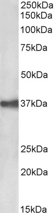
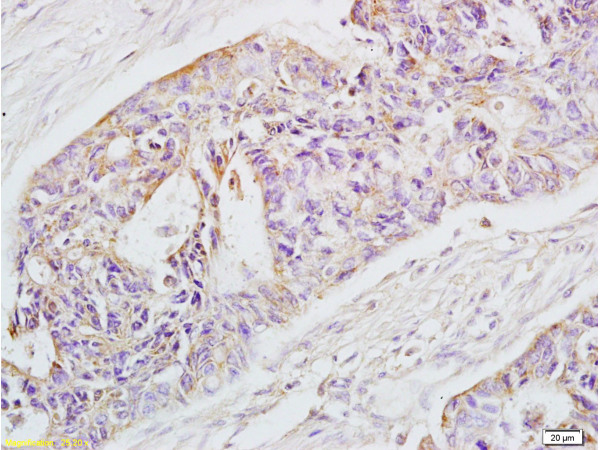
- Application Supplier NoteThe antibody is useful in immunohistochemistry and immunoblotting. MOC-31 react with antigens detectable in cryostat section. For use on frozen tissue and paraffin tissue after antigen retrieval. Using the antigen retrieval techniques this antibody discriminates between cells which originate from mesothelium and epithelium. General procedure to perform cryostat sectioning and immunostaining: Cryostat sectioning and immunostaining is done as described by de Leij et al. Sections (about 6 micron thick) are cut in a cryostat at -20°C and placed on glass slides. After drying at RT and fixation in acetone (water-free), the sections are washed 2-3 times in PBS. Subsequently (after drying the glass area around the sections) 25 ul of undiluted monoclonal antibody preparation is applied to the wet sections. After incubation for 45 mins. in a humidified atmosphere, the sections are washed again in PBS (2-3 times) and the second step reagent (an appropriately diluted HRPO-conjugated, anti mouse Ig preparation, supplemented with 1% human serum) is applied to the wet sections. After incubation for an additional 20 mins., the sections are washed again in PBS (2-3) times and the staining reaction is performed with 3-aminoethylcarbazole (10 mg dissolved in 2.5 ml dimethylformamide, subsequently 0.05 M acetate buffer, pH 4.9, is added to a total volume of 50 ml, after which the solution is filtrated and H2O2 is added to a final concentration of about 0.03%). A positive reaction is indicated by red deposit. The nuclei of the cells present in the sections are counterstained with Mayers hematoxylin to obtain a good histological picture. Working dilution: approx 1:20.. IHC, IHC-Fr, IHC-P (1:100), WB The antibody is useful in immunohistochemistry and immunoblotting. MOC-31 react with antigens detectable in cryostat section. For use on frozen tissue and paraffin tissue after antigen retrieval. Using the antigen retrieval techniques this antibody discriminates between cells which originate from mesothelium and epithelium. General procedure to perform cryostat sectioning and immunostaining: Cryostat sectioning and immunostaining is done as described by de Leij et al. Sections (about 6 micron thick) are cut in a cryostat at -20°C and placed on glass slides. After drying at RT and fixation in acetone (water-free), the sections are washed 2-3 times in PBS. Subsequently (after drying the glass area around the sections) 25 ul of undiluted monoclonal antibody preparation is applied to the wet sections. After incubation for 45 mins. in a humidified atmosphere, the sections are washed again in PBS (2-3 times) and the second step reagent (an appropriately diluted HRPO-conjugated, anti mouse Ig preparation, supplemented with 1% human serum) is applied to the wet sections. After incubation for an additional 20 mins., the sections are washed again in PBS (2-3) times and the staining reaction is performed with 3-aminoethylcarbazole (10 mg dissolved in 2.5 ml dimethylformamide, subsequently 0.05 M acetate buffer, pH 4.9, is added to a total volume of 50 ml, after which the solution is filtrated and H2O2 is added to a final concentration of about 0.03%). A positive reaction is indicated by red deposit. The nuclei of the cells present in the sections are counterstained with Mayers hematoxylin to obtain a good histological picture. Working dilution: approx 1:20.
- ApplicationsWestern Blot, ImmunoHistoChemistry, ImmunoHistoChemistry Frozen, ImmunoHistoChemistry Paraffin
- CertificationResearch Use Only
- ClonalityMonoclonal
- Clone IDMOC-31
- ConjugateUnconjugated
- Gene ID4072
- Target nameEPCAM
- Target descriptionepithelial cell adhesion molecule
- Target synonymsBer-Ep4, BerEp4, DIAR5, EGP-2, EGP314, EGP40, ESA, HNPCC8, KS1/4, KSA, LYNCH8, M4S1, MIC18, MK-1, MOC-31, TACSTD1, TROP1, epithelial cell adhesion molecule, adenocarcinoma-associated antigen, cell surface glycoprotein Trop-1, epithelial glycoprotein 314, human epithelial glycoprotein-2, major gastrointestinal tumor-associated protein GA733-2, membrane component, chromosome 4, surface marker (35kD glycoprotein), trophoblast cell surface antigen 1, tumor-associated calcium signal transducer 1
- HostMouse
- IsotypeIgG1
- ReactivityHuman
- Storage Instruction2°C to 8°C
- UNSPSC41116161
Related products
Product group Antibodies

Anti-EpCAM [AUA1]Ab00609-1.1
ApplicationsFlow Cytometry, ImmunoFluorescence, ELISA, ImmunoHistoChemistry
ReactivityHuman
TargetEPCAM
- SizePrice
Product group Antibodies
Anti-EPCAM Antibody144-01177
ApplicationsWestern Blot, ImmunoHistoChemistry
ReactivityHuman, Mouse
TargetEPCAM
- SizePrice
![EpCAM antibody [N3C3] detects EpCAM protein at cell membrane and cell junction by immunofluorescent analysis. Sample: HCT116 cells were fixed in ice-cold MeOH for 5 min. Green: EpCAM stained by EpCAM antibody [N3C3] (GTX113091) diluted at 1:500. Blue: Fluoroshield with DAPI (GTX30920).](https://www.genetex.com/upload/website/prouct_img/normal/GTX113091/GTX113091_44265_20220624_ICC_IF_22062919_816.webp)
Product group Antibodies
References
EpCAM antibody [N3C3]GTX113091
ApplicationsImmunoFluorescence, Western Blot, ImmunoCytoChemistry, ImmunoHistoChemistry, ImmunoHistoChemistry Paraffin
ReactivityHuman, Mouse
TargetEPCAM
- SizePrice
Product group Antibodies
Anti-EpCAM AntibodyA85204
ApplicationsWestern Blot, ELISA
ReactivityHuman
- SizePrice
Product group Antibodies
Epcam Polyclonal AntibodyCAC11319
ApplicationsImmunoFluorescence, Western Blot, ELISA, ImmunoHistoChemistry
ReactivityRat
TargetEPCAM
- SizePrice
Product group Antibodies
References
EpCAM Polyclonal AntibodyBS-1513R
ApplicationsFlow Cytometry, ImmunoFluorescence, Western Blot, ELISA, ImmunoCytoChemistry, ImmunoHistoChemistry, ImmunoHistoChemistry Frozen, ImmunoHistoChemistry Paraffin
ReactivityHuman, Mouse, Rat
TargetEPCAM
- SizePrice
Product group Antibodies

ApplicationsImmunoHistoChemistry, ImmunoHistoChemistry Frozen, ImmunoHistoChemistry Paraffin
ReactivityHuman
TargetEPCAM
- SizePrice