

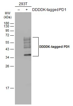
Non-transfected (–) and transfected (+) 293T whole cell extracts (30 μg) were separated by 12% SDS-PAGE, and the membrane was blotted with PD1 antibody (GTX128436) diluted at 1:5000. The HRP-conjugated anti-rabbit IgG antibody (GTX213110-01) was used to detect the primary antibody.
Non-transfected (–) and transfected (+) 293T whole cell extracts (30 μg) were separated by 12% SDS-PAGE, and the membrane was blotted with PD1 antibody (GTX128436) diluted at 1:5000. The HRP-conjugated anti-rabbit IgG antibody (GTX213110-01) was used to detect the primary antibody.
PD1 antibody
GTX128436
ApplicationsWestern Blot
Product group Antibodies
ReactivityHuman
TargetPDCD1
Overview
- SupplierGeneTex
- Product NamePD1 antibody
- Delivery Days Customer9
- Application Supplier NoteWB: 1:500-1:10000. *Optimal dilutions/concentrations should be determined by the researcher.Not tested in other applications.
- ApplicationsWestern Blot
- CertificationResearch Use Only
- ClonalityPolyclonal
- Concentration1 mg/ml
- ConjugateUnconjugated
- Gene ID5133
- Target namePDCD1
- Target descriptionprogrammed cell death 1
- Target synonymsADMIO4, AIMTBS, CD279, PD-1, PD1, SLEB2, hPD-1, hPD-l, hSLE1, programmed cell death protein 1, programmed cell death 1 protein, protein PD-1, systemic lupus erythematosus susceptibility 2
- HostRabbit
- IsotypeIgG
- Protein IDQ15116
- Protein NameProgrammed cell death protein 1
- Scientific DescriptionThis gene encodes a cell surface membrane protein of the immunoglobulin superfamily. This protein is expressed in pro-B-cells and is thought to play a role in their differentiation. In mice, expression of this gene is induced in the thymus when anti-CD3 antibodies are injected and large numbers of thymocytes undergo apoptosis. Mice deficient for this gene bred on a BALB/c background developed dilated cardiomyopathy and died from congestive heart failure. These studies suggest that this gene product may also be important in T cell function and contribute to the prevention of autoimmune diseases. [provided by RefSeq]
- ReactivityHuman
- Storage Instruction-20°C or -80°C,2°C to 8°C
- UNSPSC41116161
Datasheet
Related products
Product group Antibodies
PDCD1 AntibodyCSB-PA483633
ApplicationsELISA, ImmunoHistoChemistry
ReactivityHuman
TargetPDCD1
- SizePrice
Product group Antibodies
ApplicationsFlow Cytometry, ImmunoPrecipitation, ImmunoHistoChemistry
ReactivityHuman
TargetPDCD1
- SizePrice

Product group Antibodies
Anti-PDCD1 AntibodyAMAB91197
ApplicationsWestern Blot, ImmunoHistoChemistry
ReactivityHuman
TargetPDCD1
- SizePrice
Product group Antibodies

Anti-PD-1 [5C4.B8 (Nivolumab)]Ab00791-1.1
ApplicationsFlow Cytometry, ImmunoHistoChemistry, Neutralisation/Blocking, Other Application
ReactivityHuman, Monkey
TargetPDCD1
- SizePrice
Product group Antibodies
Anti-PDCD1 AntibodyA28569
ApplicationsWestern Blot
ReactivityHuman, Mouse, Rat
- SizePrice
Product group Antibodies

ApplicationsFlow Cytometry, ImmunoHistoChemistry
ReactivityMouse
TargetPDCD1
- SizePrice
Product group Antibodies
Anti-PD1/PDCD1 Antibody Picoband(r)RP1039-CARRIER-FREE
ApplicationsImmunoHistoChemistry
ReactivityHuman
TargetPDCD1
- SizePrice
Product group Antibodies
References
PD-1 Polyclonal AntibodyBS-1867R
ApplicationsFlow Cytometry, ImmunoFluorescence, Western Blot, ELISA, ImmunoCytoChemistry, ImmunoHistoChemistry, ImmunoHistoChemistry Frozen, ImmunoHistoChemistry Paraffin
ReactivityHuman, Mouse, Rat
TargetPDCD1
- SizePrice
Product group Antibodies
PD1 antibodyGTX128435
ApplicationsWestern Blot, ImmunoHistoChemistry, ImmunoHistoChemistry Paraffin
ReactivityHuman, Mouse
TargetPDCD1
- SizePrice