

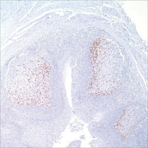
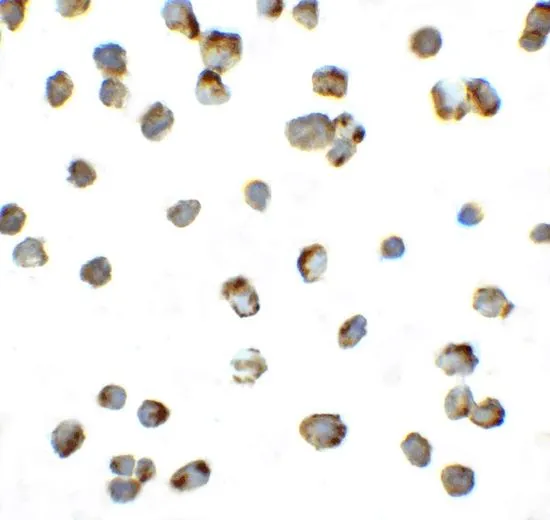
IHC-P analysis of human tonsil tissue using GTX20256 PD1 antibody [NAT105].
IHC-P analysis of human tonsil tissue using GTX20256 PD1 antibody [NAT105].
PD1 antibody [NAT105]
GTX20256
ApplicationsFlow Cytometry, ImmunoFluorescence, ImmunoPrecipitation, Western Blot, ImmunoCytoChemistry, ImmunoHistoChemistry, ImmunoHistoChemistry Frozen, ImmunoHistoChemistry Paraffin
Product group Antibodies
ReactivityHuman, Mouse
TargetPDCD1
Overview
- SupplierGeneTex
- Product NamePD1 antibody [NAT105]
- Delivery Days Customer9
- Application Supplier NoteIHC-P: 1:25-100. *Optimal dilutions/concentrations should be determined by the researcher.Not tested in other applications.
- ApplicationsFlow Cytometry, ImmunoFluorescence, ImmunoPrecipitation, Western Blot, ImmunoCytoChemistry, ImmunoHistoChemistry, ImmunoHistoChemistry Frozen, ImmunoHistoChemistry Paraffin
- CertificationResearch Use Only
- ClonalityMonoclonal
- Clone IDNAT105
- ConjugateUnconjugated
- Gene ID5133
- Target namePDCD1
- Target descriptionprogrammed cell death 1
- Target synonymsADMIO4, AIMTBS, CD279, PD-1, PD1, SLEB2, hPD-1, hPD-l, hSLE1, programmed cell death protein 1, programmed cell death 1 protein, protein PD-1, systemic lupus erythematosus susceptibility 2
- HostMouse
- IsotypeIgG1
- Protein IDQ15116
- Protein NameProgrammed cell death protein 1
- Scientific DescriptionThis gene encodes a cell surface membrane protein of the immunoglobulin superfamily. This protein is expressed in pro-B-cells and is thought to play a role in their differentiation. In mice, expression of this gene is induced in the thymus when anti-CD3 antibodies are injected and large numbers of thymocytes undergo apoptosis. Mice deficient for this gene bred on a BALB/c background developed dilated cardiomyopathy and died from congestive heart failure. These studies suggest that this gene product may also be important in T cell function and contribute to the prevention of autoimmune diseases. [provided by RefSeq, Jul 2008]
- ReactivityHuman, Mouse
- Storage Instruction-20°C or -80°C,2°C to 8°C
- UNSPSC12352203
References
- Zhang S, Minaguchi T, Xu C, et al. PD-L1 and CD4 are independent prognostic factors for overall survival in endometrial carcinomas. BMC Cancer. 2020,20(1):127. doi: 10.1186/s12885-020-6545-9Read this paper
- Paulsen EE, Kilvaer TK, Khanehkenari MR, et al. Assessing PDL-1 and PD-1 in Non-Small Cell Lung Cancer: A Novel Immunoscore Approach. Clin Lung Cancer. 2017,18(2):220-233.e8. doi: 10.1016/j.cllc.2016.09.009Read this paper
- Kaewkangsadan V, Verma C, Eremin JM, et al. Crucial Contributions by T Lymphocytes (Effector, Regulatory, and Checkpoint Inhibitor) and Cytokines (TH1, TH2, and TH17) to a Pathological Complete Response Induced by Neoadjuvant Chemotherapy in Women with Breast Cancer. J Immunol Res. 2016,2016:4757405.Read this paper
- Kammerer-Jacquet SF, Crouzet L, Brunot A, et al. Independent association of PD-L1 expression with noninactivated VHL clear cell renal cell carcinoma-A finding with therapeutic potential. Int J Cancer. 2017,140(1):142-148. doi: 10.1002/ijc.30429Read this paper
- Li CW, Lim SO, Xia W, et al. Glycosylation and stabilization of programmed death ligand-1 suppresses T-cell activity. Nat Commun. 2016,7:12632. doi: 10.1038/ncomms12632Read this paper
- Dronca RS, Liu X, Harrington SM, et al. T cell Bim levels reflect responses to anti-PD-1 cancer therapy. JCI Insight. 2016,1(6):pii: e86014.Read this paper
- Bastman JJ, Serracino HS, Zhu Y, et al. Tumor-Infiltrating T Cells and the PD-1 Checkpoint Pathway in Advanced Differentiated and Anaplastic Thyroid Cancer. J Clin Endocrinol Metab. 2016,101(7):2863-73. doi: 10.1210/jc.2015-4227Read this paper
- Mishra V, Schuetz H, Haorah J. Differential induction of PD-1/PD-L1 in Neuroimmune cells by drug of abuse. Int J Physiol Pathophysiol Pharmacol. 2015,7(2):87-97.Read this paper
- Joseph RW, Cappel M, Goedjen B, et al. Lichenoid dermatitis in three patients with metastatic melanoma treated with anti-PD-1 therapy. Cancer Immunol Res. 2015,3(1):18-22. doi: 10.1158/2326-6066.CIR-14-0134Read this paper
- Szabo K, Papp G, Dezso B, et al. The histopathology of labial salivary glands in primary Sjögren's syndrome: focusing on follicular helper T cells in the inflammatory infiltrates. Mediators Inflamm. 2014,2014:631787. doi: 10.1155/2014/631787Read this paper
Datasheet
Related products
Product group Antibodies
Pdcd1 Polyclonal AntibodyCAC07144
ApplicationsImmunoFluorescence, Western Blot, ELISA, ImmunoHistoChemistry
ReactivityMouse
TargetPDCD1
- SizePrice
Product group Antibodies
ApplicationsFlow Cytometry, ImmunoPrecipitation, ImmunoHistoChemistry
ReactivityHuman
TargetPDCD1
- SizePrice
Product group Antibodies
Anti-PDCD1 AntibodyA28569
ApplicationsWestern Blot
ReactivityHuman, Mouse, Rat
- SizePrice
Product group Antibodies
ApplicationsNeutralisation/Blocking
TargetPDCD1
- SizePrice

Product group Antibodies
Anti-PDCD1 AntibodyAMAB91197
ApplicationsWestern Blot, ImmunoHistoChemistry
ReactivityHuman
TargetPDCD1
- SizePrice
Product group Antibodies
anti-PD-1 (human), mAb (AG-IHC001)AG-20B-6020
ApplicationsELISA, ImmunoHistoChemistry
ReactivityHuman
TargetPDCD1
- SizePrice
Product group Antibodies

Anti-PD-1 [5C4.B8 (Nivolumab)]Ab00791-1.1
ApplicationsFlow Cytometry, ImmunoHistoChemistry, Neutralisation/Blocking, Other Application
ReactivityHuman, Monkey
TargetPDCD1
- SizePrice
Product group Antibodies
PD1 antibodyGTX31309
ApplicationsImmunoFluorescence, Western Blot, ELISA, ImmunoCytoChemistry, ImmunoHistoChemistry, ImmunoHistoChemistry Paraffin
ReactivityHuman, Mouse, Rat
TargetPDCD1
- SizePrice