

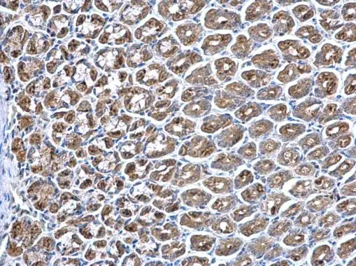
PPT1 antibody [N1C3] detects PPT1 protein at lysosome on mouse stomach by immunohistochemical analysis. Sample: Paraffin-embedded mouse stomach. PPT1 antibody [N1C3] (GTX110677) dilution: 1:500. Scale bar = M μm.
Antigen Retrieval: Trilogy? (EDTA based, pH 8.0) buffer, 15min
 antibody at 1:500 dilution.
Antigen Retrieval: Trilogy? (EDTA based, pH 8.0) buffer, 15min")
![PPT1 antibody [N1C3] detects PPT1 protein at lysosome on rat hind brain by immunohistochemical analysis. Sample: Paraffin-embedded rat hind brain. PPT1 antibody [N1C3] (GTX110677) dilution: 1:500.
Antigen Retrieval: Trilogy? (EDTA based, pH 8.0) buffer, 15min](https://www.genetex.com/upload/website/prouct_img/normal/GTX110677/GTX110677_40073_IHC_R_w_23060500_765.webp "PPT1 antibody [N1C3] detects PPT1 protein at lysosome on rat hind brain by immunohistochemical analysis. Sample: Paraffin-embedded rat hind brain. PPT1 antibody [N1C3] (GTX110677) dilution: 1:500.
Antigen Retrieval: Trilogy? (EDTA based, pH 8.0) buffer, 15min")
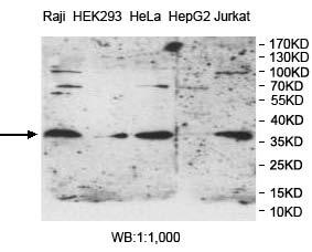
 A: Hela 10% SDS PAGE GTX110677 diluted at 1:3000")
![PPT1 antibody [N1C3] detects PPT1 protein at lysosome on mouse fore brain by immunohistochemical analysis. Sample: Paraffin-embedded mouse fore brain. PPT1 antibody [N1C3] (GTX110677) dilution: 1:500. Scale bar = M_2 μm.
Antigen Retrieval: Trilogy? (EDTA based, pH 8.0) buffer, 15min](https://www.genetex.com/upload/website/prouct_img/normal/GTX110677/GTX110677_40073_IHC_M_2_w_23060500_710.webp "PPT1 antibody [N1C3] detects PPT1 protein at lysosome on mouse fore brain by immunohistochemical analysis. Sample: Paraffin-embedded mouse fore brain. PPT1 antibody [N1C3] (GTX110677) dilution: 1:500. Scale bar = M_2 μm.
Antigen Retrieval: Trilogy? (EDTA based, pH 8.0) buffer, 15min")
PPT1 antibody [N1C3] detects PPT1 protein at lysosome on mouse stomach by immunohistochemical analysis. Sample: Paraffin-embedded mouse stomach. PPT1 antibody [N1C3] (GTX110677) dilution: 1:500. Scale bar = M μm.
Antigen Retrieval: Trilogy? (EDTA based, pH 8.0) buffer, 15min
PPT1 antibody [N1C3]
GTX110677
ApplicationsImmunoFluorescence, Western Blot, ImmunoCytoChemistry, ImmunoHistoChemistry, ImmunoHistoChemistry Paraffin
Product group Antibodies
ReactivityHuman, Mouse, Rat
TargetPPT1
Overview
- SupplierGeneTex
- Product NamePPT1 antibody [N1C3]
- Delivery Days Customer9
- Application Supplier NoteWB: 1:500-1:3000. IHC-P: 1:100-1:1000. *Optimal dilutions/concentrations should be determined by the researcher.Not tested in other applications.
- ApplicationsImmunoFluorescence, Western Blot, ImmunoCytoChemistry, ImmunoHistoChemistry, ImmunoHistoChemistry Paraffin
- CertificationResearch Use Only
- ClonalityPolyclonal
- Concentration1 mg/ml
- ConjugateUnconjugated
- Gene ID5538
- Target namePPT1
- Target descriptionpalmitoyl-protein thioesterase 1
- Target synonymsCLN1, INCL, PPT, palmitoyl-protein thioesterase 1, ceroid-palmitoyl-palmitoyl-protein thioesterase 1, palmitoyl-protein hydrolase 1
- HostRabbit
- IsotypeIgG
- Protein IDP50897
- Protein NamePalmitoyl-protein thioesterase 1
- Scientific DescriptionThe protein encoded by this gene is a small glycoprotein involved in the catabolism of lipid-modified proteins during lysosomal degradation. The encoded enzyme removes thioester-linked fatty acyl groups such as palmitate from cysteine residues. Defects in this gene are a cause of infantile neuronal ceroid lipofuscinosis 1 (CLN1, or INCL) and neuronal ceroid lipofuscinosis 4 (CLN4). Two transcript variants encoding different isoforms have been found for this gene.
- ReactivityHuman, Mouse, Rat
- Storage Instruction-20°C or -80°C,2°C to 8°C
- UNSPSC41116161
Datasheet
Related products
Product group Antibodies
Anti-PPT1 AntibodyA48574
ApplicationsWestern Blot, ELISA, ImmunoHistoChemistry
ReactivityHuman, Mouse, Rat
- SizePrice
Product group Antibodies
Anti-PPT1 Antibody144-63441
ApplicationsWestern Blot
ReactivityHuman, Mouse, Rat
TargetPPT1
- SizePrice
Product group Antibodies
PPT1 Polyclonal AntibodyBS-6619R
ApplicationsImmunoFluorescence, Western Blot, ELISA, ImmunoCytoChemistry, ImmunoHistoChemistry, ImmunoHistoChemistry Frozen, ImmunoHistoChemistry Paraffin
ReactivityBovine, Canine, Chicken, Equine, Human, Mouse, Porcine, Rabbit, Rat, Sheep
TargetPPT1
- SizePrice
Product group Antibodies
PPT1 Polyclonal AntibodyCAC14868
ApplicationsWestern Blot, ELISA, ImmunoHistoChemistry
TargetPPT1
- SizePrice
Product group Antibodies
PPT1 AntibodyCSB-PA018587LA01HU
ApplicationsWestern Blot, ELISA, ImmunoHistoChemistry
ReactivityHuman
TargetPPT1
- SizePrice
Product group Antibodies

PPT1 / CLN1 Antibody (N-Terminus)LS-C358836
ApplicationsWestern Blot, ImmunoHistoChemistry, ImmunoHistoChemistry Paraffin
ReactivityBovine, Human, Monkey
TargetPPT1
- SizePrice

Product group Antibodies
Anti-PPT1 AntibodyHPA021546
ApplicationsImmunoHistoChemistry
ReactivityHuman
TargetPPT1
- SizePrice

Product group Antibodies
Anti-PPT1 Antibody Picoband(r)PB9781-CARRIER-FREE
ApplicationsFlow Cytometry, Western Blot, ImmunoCytoChemistry, ImmunoHistoChemistry, ImmunoHistoChemistry Frozen
ReactivityHuman, Mouse, Rat
TargetPPT1
- SizePrice
Product group Antibodies
Anti-PPT1Y158470
ApplicationsWestern Blot, ELISA, ImmunoHistoChemistry
ReactivityHuman
- SizePrice