

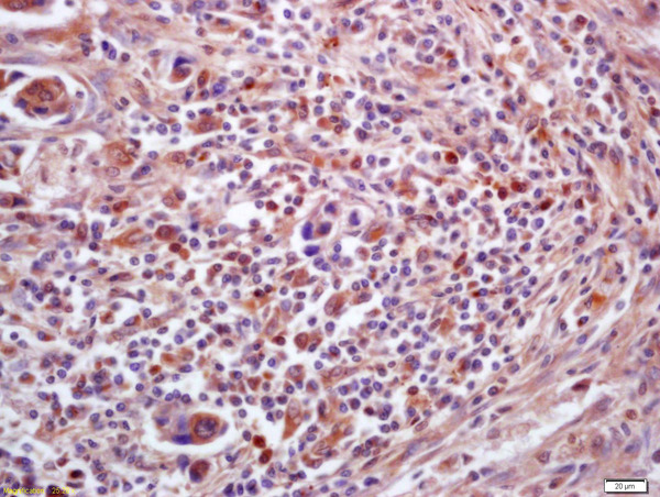
Formalin-fixed and paraffin embedded human lung carcinoma labeled with Anti-PPT1 Polyclonal Antibody, Unconjugated (bs-6619R) at 1:200 followed by conjugation to the secondary antibody and DAB staining
 at 1:200 followed by conjugation to the secondary antibody and DAB staining")
Formalin-fixed and paraffin embedded human lung carcinoma labeled with Anti-PPT1 Polyclonal Antibody, Unconjugated (bs-6619R) at 1:200 followed by conjugation to the secondary antibody and DAB staining
PPT1 Polyclonal Antibody
BS-6619R
ApplicationsImmunoFluorescence, Western Blot, ELISA, ImmunoCytoChemistry, ImmunoHistoChemistry, ImmunoHistoChemistry Frozen, ImmunoHistoChemistry Paraffin
Product group Antibodies
ReactivityBovine, Canine, Chicken, Equine, Human, Mouse, Porcine, Rabbit, Rat, Sheep
TargetPPT1
Overview
- SupplierBioss
- Product NamePPT1 Polyclonal Antibody
- Delivery Days Customer16
- ApplicationsImmunoFluorescence, Western Blot, ELISA, ImmunoCytoChemistry, ImmunoHistoChemistry, ImmunoHistoChemistry Frozen, ImmunoHistoChemistry Paraffin
- Applications SupplierWB(1:300-5000), ELISA(1:500-1000), IHC-P(1:200-400), IHC-F(1:100-500), IF(IHC-P)(1:50-200), IF(IHC-F)(1:50-200), IF(ICC)(1:50-200)
- CertificationResearch Use Only
- ClonalityPolyclonal
- Concentration1 ug/ul
- ConjugateUnconjugated
- Gene ID5538
- Target namePPT1
- Target descriptionpalmitoyl-protein thioesterase 1
- Target synonymsCLN1, INCL, PPT, palmitoyl-protein thioesterase 1, ceroid-palmitoyl-palmitoyl-protein thioesterase 1, palmitoyl-protein hydrolase 1
- HostRabbit
- IsotypeIgG
- Protein IDP50897
- Protein NamePalmitoyl-protein thioesterase 1
- ReactivityBovine, Canine, Chicken, Equine, Human, Mouse, Porcine, Rabbit, Rat, Sheep
- Storage Instruction-20°C
- UNSPSC41116161
Datasheet
Related products
Product group Antibodies
Anti-PPT1 AntibodyA48574
ApplicationsWestern Blot, ELISA, ImmunoHistoChemistry
ReactivityHuman, Mouse, Rat
- SizePrice
Product group Antibodies
Anti-PPT1 Antibody144-63441
ApplicationsWestern Blot
ReactivityHuman, Mouse, Rat
TargetPPT1
- SizePrice
Product group Antibodies
PPT1 Polyclonal AntibodyCAC14868
ApplicationsWestern Blot, ELISA, ImmunoHistoChemistry
TargetPPT1
- SizePrice
Product group Antibodies
PPT1 AntibodyCSB-PA018587LA01HU
ApplicationsWestern Blot, ELISA, ImmunoHistoChemistry
ReactivityHuman
TargetPPT1
- SizePrice
Product group Antibodies

PPT1 / CLN1 Antibody (N-Terminus)LS-C358836
ApplicationsWestern Blot, ImmunoHistoChemistry, ImmunoHistoChemistry Paraffin
ReactivityBovine, Human, Monkey
TargetPPT1
- SizePrice

Product group Antibodies
Anti-PPT1 AntibodyHPA021546
ApplicationsImmunoHistoChemistry
ReactivityHuman
TargetPPT1
- SizePrice
![PPT1 antibody [N1C3] detects PPT1 protein at lysosome on mouse stomach by immunohistochemical analysis. Sample: Paraffin-embedded mouse stomach. PPT1 antibody [N1C3] (GTX110677) dilution: 1:500. Scale bar = M μm.
Antigen Retrieval: Trilogy? (EDTA based, pH 8.0) buffer, 15min](https://www.genetex.com/upload/website/prouct_img/normal/GTX110677/GTX110677_40073_IHC_M_w_23060500_974.webp)
Product group Antibodies
PPT1 antibody [N1C3]GTX110677
ApplicationsImmunoFluorescence, Western Blot, ImmunoCytoChemistry, ImmunoHistoChemistry, ImmunoHistoChemistry Paraffin
ReactivityHuman, Mouse, Rat
TargetPPT1
- SizePrice

Product group Antibodies
Anti-PPT1 Antibody Picoband(r)PB9781-CARRIER-FREE
ApplicationsFlow Cytometry, Western Blot, ImmunoCytoChemistry, ImmunoHistoChemistry, ImmunoHistoChemistry Frozen
ReactivityHuman, Mouse, Rat
TargetPPT1
- SizePrice
Product group Antibodies
Anti-PPT1Y158470
ApplicationsWestern Blot, ELISA, ImmunoHistoChemistry
ReactivityHuman
- SizePrice