

Primary Antibodies
Product group Antibodies
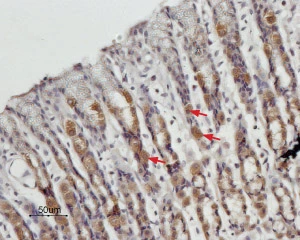
Orexin Receptor 2 antibodyGTX54848
ApplicationsImmunoFluorescence, Western Blot, ImmunoCytoChemistry, ImmunoHistoChemistry, ImmunoHistoChemistry Frozen
ReactivityHuman, Mouse, Rat
TargetHcrtr2
- SizePrice
Product group Antibodies
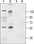
P2X1 antibodyGTX54849
ApplicationsFlow Cytometry, ImmunoFluorescence, ImmunoPrecipitation, Western Blot, ImmunoCytoChemistry, ImmunoHistoChemistry, ImmunoHistoChemistry Frozen
ReactivityHuman, Mouse, Rat
TargetP2rx1
- SizePrice
Product group Antibodies
References
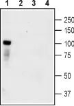
P2X4 antibodyGTX54851
ApplicationsFlow Cytometry, ImmunoFluorescence, ImmunoPrecipitation, Western Blot, ImmunoCytoChemistry, ImmunoHistoChemistry, ImmunoHistoChemistry Frozen, ImmunoHistoChemistry Paraffin
ReactivityHuman, Mouse, Rat
TargetP2rx4
- SizePrice

Product group Antibodies
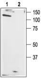
PICK1 antibodyGTX54853
ApplicationsWestern Blot, ImmunoHistoChemistry, ImmunoHistoChemistry Frozen
ReactivityHuman, Mouse, Rat
TargetPick1
- SizePrice
Product group Antibodies
References
Sortilin 1 antibodyGTX54854
ApplicationsFlow Cytometry, Western Blot, ImmunoHistoChemistry, ImmunoHistoChemistry Frozen
ReactivityHuman, Mouse, Rat
TargetSORT1
- SizePrice
Product group Antibodies
References
STIM1 antibodyGTX54855
ApplicationsFlow Cytometry, ImmunoFluorescence, ImmunoPrecipitation, Western Blot, ImmunoCytoChemistry, ImmunoHistoChemistry, ImmunoHistoChemistry Paraffin, Other Application
ReactivityHuman, Mouse, Rat
TargetSTIM1
- SizePrice
Product group Antibodies
References
TrkA antibodyGTX54856
ApplicationsFlow Cytometry, ImmunoFluorescence, Western Blot, ImmunoCytoChemistry, ImmunoHistoChemistry, ImmunoHistoChemistry Frozen, Other Application
ReactivityHuman, Mouse, Rat
TargetNtrk1
- SizePrice
Product group Antibodies
References
TrkB antibodyGTX54857
ApplicationsFlow Cytometry, ImmunoFluorescence, Western Blot, ImmunoCytoChemistry, ImmunoHistoChemistry, ImmunoHistoChemistry Frozen, Other Application
ReactivityHuman, Mouse, Rat
TargetNtrk2
- SizePrice
Product group Antibodies
References
TrkC antibodyGTX54858
ApplicationsImmunoFluorescence, Western Blot, ImmunoCytoChemistry, ImmunoHistoChemistry, ImmunoHistoChemistry Frozen, ImmunoHistoChemistry Paraffin, Other Application
ReactivityHuman, Mouse, Rat
TargetNtrk3
- SizePrice
Product group Antibodies
References
TRPC1 antibodyGTX54859
ApplicationsImmunoFluorescence, ImmunoPrecipitation, Western Blot, ImmunoCytoChemistry, ImmunoHistoChemistry, ImmunoHistoChemistry Frozen
ReactivityHuman, Mouse, Rat
TargetTRPC1
- SizePrice
Product group Antibodies
References
TRPC3 antibodyGTX54860
ApplicationsFlow Cytometry, ImmunoFluorescence, ImmunoPrecipitation, Western Blot, ImmunoCytoChemistry, ImmunoHistoChemistry, ImmunoHistoChemistry Frozen, ImmunoHistoChemistry Paraffin
ReactivityHuman, Mouse, Rat
TargetTrpc3
- SizePrice
Product group Antibodies
References
TRPC5 antibodyGTX54861
ApplicationsImmunoFluorescence, ImmunoPrecipitation, Western Blot, ImmunoCytoChemistry, ImmunoHistoChemistry, ImmunoHistoChemistry Frozen
ReactivityHuman, Mouse, Rat
TargetTRPC5
- SizePrice
Didn't find what you were looking for?
Search through our product groups to find the right product
Back to overview