

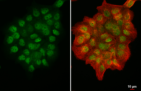
PRMT5 antibody [N3C3] detects PRMT5 protein at nucleus by immunofluorescent analysis. Sample: A431 cells were fixed in 4% paraformaldehyde at RT for 15 min. Green: PRMT5 stained by PRMT5 antibody [N3C3] (GTX115409) diluted at 1:500. Red: alpha Tubulin, a cytoskeleton marker, stained by alpha Tubulin antibody [GT114] (GTX628802) diluted at 1:1000.
 antibody at 1:100 dilution.
Antigen Retrieval: Trilogy? (EDTA based, pH 8.0) buffer, 15min")
![PRMT5 antibody [N3C3] detects PRMT5 protein at cytosol and nucleus on mouse colon by immunohistochemical analysis. Sample: Paraffin-embedded mouse colon. PRMT5 antibody [N3C3] (GTX115409) dilution: 1:500.
Antigen Retrieval: Trilogy? (EDTA based, pH 8.0) buffer, 15min](https://www.genetex.com/upload/website/prouct_img/normal/GTX115409/GTX115409_40177_IHC_M_2_w_23060519_792.webp "PRMT5 antibody [N3C3] detects PRMT5 protein at cytosol and nucleus on mouse colon by immunohistochemical analysis. Sample: Paraffin-embedded mouse colon. PRMT5 antibody [N3C3] (GTX115409) dilution: 1:500.
Antigen Retrieval: Trilogy? (EDTA based, pH 8.0) buffer, 15min")
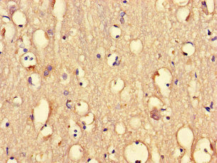
![PRMT5 antibody [N3C3] detects PRMT5 protein at cytoplasm and nucleus by immunohistochemical analysis. Sample: Paraffin-embedded mouse testis. PRMT5 stained by PRMT5 antibody [N3C3] (GTX115409) diluted at 1:2500. Antigen Retrieval: Citrate buffer, pH 6.0, 15 min](https://www.genetex.com/upload/website/prouct_img/normal/GTX115409/GTX115409_44384_20211119_IHC-P_M_w_23060519_236.webp "PRMT5 antibody [N3C3] detects PRMT5 protein at cytoplasm and nucleus by immunohistochemical analysis. Sample: Paraffin-embedded mouse testis. PRMT5 stained by PRMT5 antibody [N3C3] (GTX115409) diluted at 1:2500. Antigen Retrieval: Citrate buffer, pH 6.0, 15 min")
![PRMT5 antibody [N3C3] detects PRMT5 protein at cytosol and nucleus on mouse colon by immunohistochemical analysis. Sample: Paraffin-embedded mouse colon. PRMT5 antibody [N3C3] (GTX115409) dilution: 1:500.
Antigen Retrieval: Trilogy? (EDTA based, pH 8.0) buffer, 15min](https://www.genetex.com/upload/website/prouct_img/normal/GTX115409/GTX115409_40177_IHC_M_w_23060519_466.webp "PRMT5 antibody [N3C3] detects PRMT5 protein at cytosol and nucleus on mouse colon by immunohistochemical analysis. Sample: Paraffin-embedded mouse colon. PRMT5 antibody [N3C3] (GTX115409) dilution: 1:500.
Antigen Retrieval: Trilogy? (EDTA based, pH 8.0) buffer, 15min")
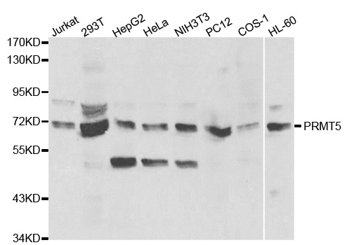
![Various whole cell extracts (30 μg) were separated by 7.5% SDS-PAGE, and the membrane was blotted with PRMT5 antibody [N3C3] (GTX115409) diluted at 1:1000. The HRP-conjugated anti-rabbit IgG antibody (GTX213110-01) was used to detect the primary antibody.](https://www.genetex.com/upload/website/prouct_img/normal/GTX115409/GTX115409_44384_20210730_WB_w_23060519_815.webp "Various whole cell extracts (30 μg) were separated by 7.5% SDS-PAGE, and the membrane was blotted with PRMT5 antibody [N3C3] (GTX115409) diluted at 1:1000. The HRP-conjugated anti-rabbit IgG antibody (GTX213110-01) was used to detect the primary antibody.")
PRMT5 antibody [N3C3] detects PRMT5 protein at nucleus by immunofluorescent analysis. Sample: A431 cells were fixed in 4% paraformaldehyde at RT for 15 min. Green: PRMT5 stained by PRMT5 antibody [N3C3] (GTX115409) diluted at 1:500. Red: alpha Tubulin, a cytoskeleton marker, stained by alpha Tubulin antibody [GT114] (GTX628802) diluted at 1:1000.
PRMT5 antibody [N3C3]
GTX115409
ApplicationsImmunoFluorescence, Western Blot, ImmunoCytoChemistry, ImmunoHistoChemistry, ImmunoHistoChemistry Paraffin
Product group Antibodies
ReactivityHuman, Mouse
TargetPRMT5
Overview
- SupplierGeneTex
- Product NamePRMT5 antibody [N3C3]
- Delivery Days Customer9
- Application Supplier NoteWB: 1:500-1:3000. ICC/IF: 1:100-1:1000. IHC-P: 1:100-1:1000. *Optimal dilutions/concentrations should be determined by the researcher.Not tested in other applications.
- ApplicationsImmunoFluorescence, Western Blot, ImmunoCytoChemistry, ImmunoHistoChemistry, ImmunoHistoChemistry Paraffin
- CertificationResearch Use Only
- ClonalityPolyclonal
- Concentration0.31 mg/ml
- ConjugateUnconjugated
- Gene ID10419
- Target namePRMT5
- Target descriptionprotein arginine methyltransferase 5
- Target synonymsHRMT1L5, HSL7, IBP72, JBP1, SKB1, SKB1Hs, protein arginine N-methyltransferase 5, 72 kDa ICln-binding protein, HMT1 hnRNP methyltransferase-like 5, SKB1 homolog, histone-arginine N-methyltransferase PRMT5, jak-binding protein 1, shk1 kinase-binding protein 1 homolog
- HostRabbit
- IsotypeIgG
- Protein IDO14744
- Protein NameProtein arginine N-methyltransferase 5
- Scientific DescriptionArginine methyltransferase that can both catalyze the formation of omega-N monomethylarginine (MMA) and symmetrical dimethylarginine (sDMA), with a preference for the formation of MMA. Specifically mediates the symmetrical dimethylation of arginine residues in the small nuclear ribonucleoproteins Sm D1 (SNRPD1) and Sm D3 (SNRPD3); such methylation being required for the assembly and biogenesis of snRNP core particles. Methylates SUPT5H. Mono- and dimethylates arginine residues of myelin basic protein (MBP) in vitro. Plays a role in the assembly of snRNP core particles. May play a role in cytokine-activated transduction pathways. Negatively regulates cyclin E1 promoter activity and cellular proliferation. May regulate the SUPT5H transcriptional elongation properties. May be part of a pathway that is connected to a chloride current, possibly through cytoskeletal rearrangement. Methylates histone H2A and H4 Arg-3 during germ cell development. Methylates histone H3 Arg-8, which may repress transcription. Methylates the Piwi proteins (PIWIL1, PIWIL2 and PIWIL4), methylation of Piwi proteins being required for the interaction with Tudor domain-containing proteins and subsequent localization to the meiotic nuage. Methylates RPS10.
- ReactivityHuman, Mouse
- Storage Instruction-20°C or -80°C,2°C to 8°C
- UNSPSC41116161
Datasheet
Related products
Product group Antibodies
PRMT5 AntibodyCSB-PA018734LA01HU
ApplicationsImmunoFluorescence, ELISA, ImmunoHistoChemistry
ReactivityHuman
TargetPRMT5
- SizePrice

Product group Antibodies
Anti-PRMT5 Antibody Picoband(r)A00635-1-CARRIER-FREE
ApplicationsFlow Cytometry, Western Blot, ELISA
ReactivityHuman, Mouse, Rat
TargetPRMT5
- SizePrice
Product group Antibodies

Anti-PRMT5 [RAB-C136]Ab01705-1.1
ApplicationsImmunoFluorescence, ImmunoPrecipitation, ChIP Chromatin ImmunoPrecipitation, ELISA
ReactivityHuman
TargetPRMT5
- SizePrice
Product group Antibodies
Anti-PRMT5 AntibodyA30085
ApplicationsImmunoFluorescence, ImmunoPrecipitation, Western Blot, ChIP Chromatin ImmunoPrecipitation, ImmunoHistoChemistry
ReactivityHuman, Mouse
- SizePrice
Product group Antibodies

Goat anti-PRMT5EB09534
ApplicationsWestern Blot, ELISA
ReactivityBovine, Canine, Human, Mouse, Porcine, Rat
TargetPRMT5
- SizePrice

Product group Antibodies
Anti-PRMT5 AntibodyHPA005525
ApplicationsWestern Blot, ImmunoCytoChemistry, ImmunoHistoChemistry
ReactivityHuman, Mouse, Rat
TargetPRMT5
- SizePrice
Product group Antibodies

PRMT5 AntibodyLS-C402521
ApplicationsWestern Blot, ELISA, ImmunoHistoChemistry
ReactivityHuman, Mouse, Rat
TargetPRMT5
- SizePrice
Product group Antibodies
Prmt5 Recombinant AntibodyCAC12525
ApplicationsWestern Blot, ELISA, ImmunoHistoChemistry
TargetPRMT5
- SizePrice
Product group Antibodies
PRMT5 Recombinant AntibodyBSM-60525R
ApplicationsFlow Cytometry, ImmunoFluorescence, Western Blot, ImmunoHistoChemistry, ImmunoHistoChemistry Frozen, ImmunoHistoChemistry Paraffin
ReactivityHuman
TargetPRMT5
- SizePrice