

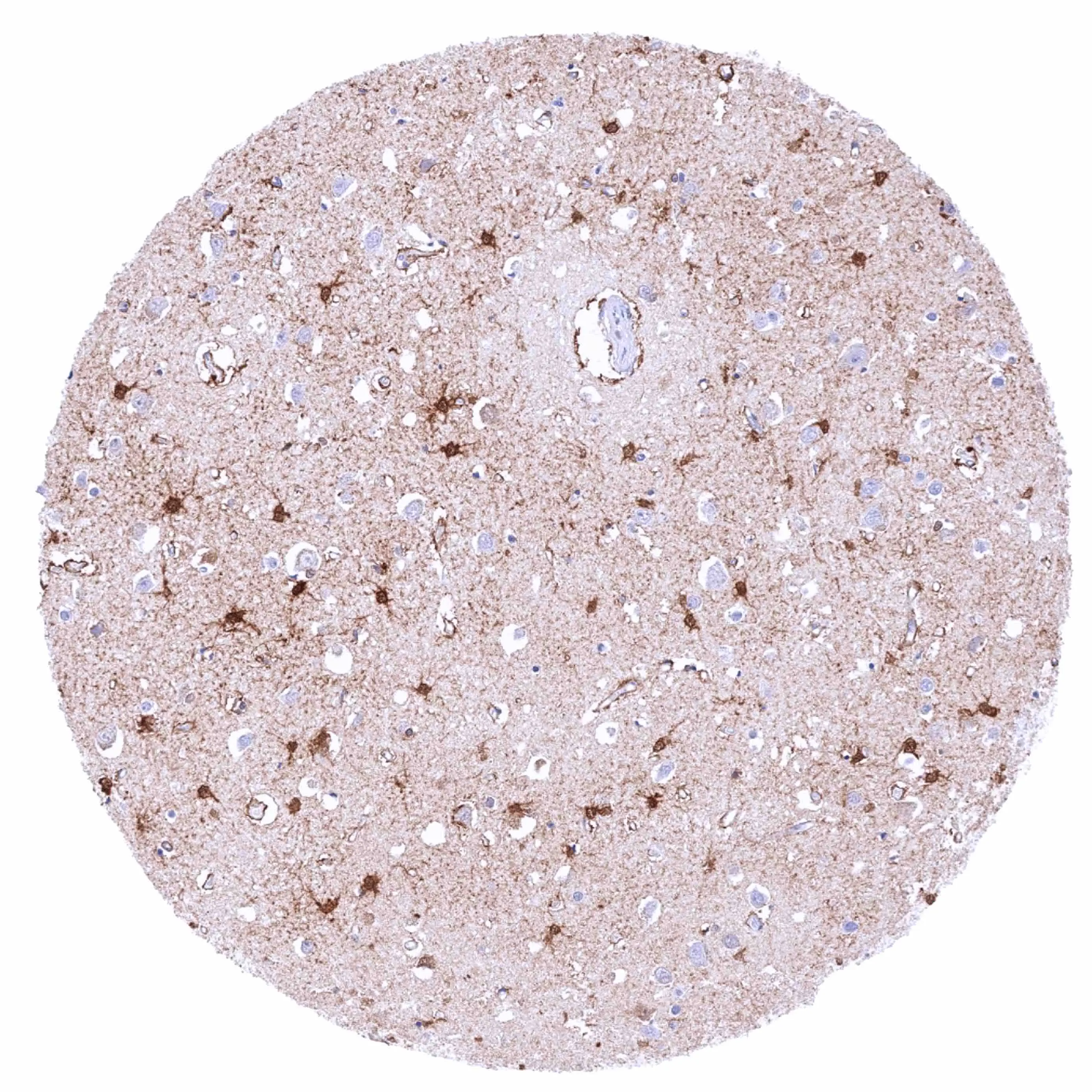
IHC-P analysis of human cerebrum (grey matter) tissue using GTX640563 PSAT1 antibody [HMV331] HistoMAX?. Strong nuclear and cytoplasmic PSAT1 staining and mainly of astrocytes.
![IHC-P analysis of human cerebellum cortex (molecular layer, Purkinje cell layer, and granule cell layer) tissue using GTX640563 PSAT1 antibody [HMV331] HistoMAX?. Nuclear and cytoplasmic PSAT1 staining is particularly strong in a subset of glial cells between the molecular and the granule cell.](https://www.genetex.com/upload/website/prouct_img/normal/GTX640563/GTX640563_20240703_IHC-P_1_24070300_324.webp "IHC-P analysis of human cerebellum cortex (molecular layer, Purkinje cell layer, and granule cell layer) tissue using GTX640563 PSAT1 antibody [HMV331] HistoMAX?. Nuclear and cytoplasmic PSAT1 staining is particularly strong in a subset of glial cells between the molecular and the granule cell.")
![IHC-P analysis of human cerebellum (white matter) tissue using GTX640563 PSAT1 antibody [HMV331] HistoMAX?. Strong nuclear and cytoplasmic PSAT1 staining of glial cells.](https://www.genetex.com/upload/website/prouct_img/normal/GTX640563/GTX640563_20240703_IHC-P_24070300_274.webp "IHC-P analysis of human cerebellum (white matter) tissue using GTX640563 PSAT1 antibody [HMV331] HistoMAX?. Strong nuclear and cytoplasmic PSAT1 staining of glial cells.")
![IHC-P analysis of human pulmonary adenocarcinoma tissue using GTX640563 PSAT1 antibody [HMV331] HistoMAX?. A moderate to A strong PSAT1 staining of most tumor cells.](https://www.genetex.com/upload/website/prouct_img/normal/GTX640563/GTX640563_20250214_IHC-P_1_25021323_701.webp "IHC-P analysis of human pulmonary adenocarcinoma tissue using GTX640563 PSAT1 antibody [HMV331] HistoMAX?. A moderate to A strong PSAT1 staining of most tumor cells.")
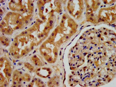
![IHC-P analysis of human kidney tissue using GTX640563 PSAT1 antibody [HMV331] HistoMAX?. A predominantly cytoplasmic PSAT1 staining of variable intensity in most tubuli.](https://www.genetex.com/upload/website/prouct_img/normal/GTX640563/GTX640563_20250214_IHC-P_25021323_712.webp "IHC-P analysis of human kidney tissue using GTX640563 PSAT1 antibody [HMV331] HistoMAX?. A predominantly cytoplasmic PSAT1 staining of variable intensity in most tubuli.")
IHC-P analysis of human cerebrum (grey matter) tissue using GTX640563 PSAT1 antibody [HMV331] HistoMAX?. Strong nuclear and cytoplasmic PSAT1 staining and mainly of astrocytes.
PSAT1 antibody [HMV331] HistoMAX(tm)
GTX640563
ApplicationsImmunoHistoChemistry, ImmunoHistoChemistry Paraffin
Product group Antibodies
ReactivityHuman
TargetPSAT1
Overview
- SupplierGeneTex
- Product NamePSAT1 antibody [HMV331] HistoMAX(tm)
- Delivery Days Customer7
- Application Supplier NoteIHC-P: 1:100-1:200. *Optimal dilutions/concentrations should be determined by the researcher.Not tested in other applications.
- ApplicationsImmunoHistoChemistry, ImmunoHistoChemistry Paraffin
- CertificationResearch Use Only
- ClonalityMonoclonal
- Clone IDHMV331
- Concentration7 ug/ml
- ConjugateUnconjugated
- Gene ID29968
- Target namePSAT1
- Target descriptionphosphoserine aminotransferase 1
- Target synonymsEPIP, NLS2, PSA, PSAT, PSATD, phosphoserine aminotransferase, endometrial progesterone-induced protein, phosphohydroxythreonine aminotransferase, phosphoserine transaminase
- HostRabbit
- IsotypeIgG
- Protein IDQ9Y617
- Protein NamePhosphoserine aminotransferase
- Scientific DescriptionThis gene encodes a member of the class-V pyridoxal-phosphate-dependent aminotransferase family. The encoded protein is a phosphoserine aminotransferase and decreased expression may be associated with schizophrenia. Mutations in this gene are also associated with phosphoserine aminotransferase deficiency. Alternative splicing results in multiple transcript variants. Pseudogenes of this gene have been defined on chromosomes 1, 3, and 8. [provided by RefSeq, Jul 2013]
- ReactivityHuman
- Storage Instruction-20°C or -80°C,2°C to 8°C
- UNSPSC41116161
Datasheet
Related products
Product group Antibodies
PSAT1 AntibodyCSB-PA018838LA01HU
ApplicationsImmunoFluorescence, ELISA, ImmunoHistoChemistry
ReactivityHuman
TargetPSAT1
- SizePrice

Product group Antibodies
Anti-PSAT1 Antibody Picoband(r)A06277-CARRIER-FREE
ApplicationsFlow Cytometry, ImmunoFluorescence, Western Blot, ELISA, ImmunoCytoChemistry
ReactivityHuman, Mouse, Rat
TargetPSAT1
- SizePrice
Product group Antibodies
Anti-PSAT1 AntibodyA31537
ApplicationsWestern Blot, ImmunoHistoChemistry
ReactivityHuman, Mouse, Rat
- SizePrice
Product group Antibodies

PSAT1 AntibodyLS-C749133
ApplicationsWestern Blot, ImmunoHistoChemistry
ReactivityHuman, Mouse, Rat
TargetPSAT1
- SizePrice

Product group Antibodies
Anti-PSAT1 AntibodyHPA042924
ApplicationsWestern Blot, ImmunoCytoChemistry
ReactivityHuman
TargetPSAT1
- SizePrice
Product group Antibodies
ApplicationsWestern Blot, ImmunoHistoChemistry
ReactivityMouse, Porcine, Rat
TargetPSAT1
- SizePrice
![Various whole cell extracts (30 μg) were separated by 10% SDS-PAGE, and the membrane was blotted with PSAT1 antibody [HL2270] (GTX638322) diluted at 1:1000. The HRP-conjugated anti-rabbit IgG antibody (GTX213110-01) was used to detect the primary antibody.](https://www.genetex.com/upload/website/prouct_img/normal/GTX638322/GTX638322_T-44970_20230317_WB_23032022_458.webp)
Product group Antibodies
PSAT1 antibody [HL2270]GTX638322
ApplicationsWestern Blot, ImmunoHistoChemistry, ImmunoHistoChemistry Paraffin
ReactivityHuman, Mouse, Rat
TargetPSAT1
- SizePrice
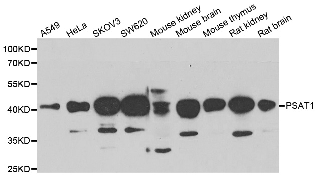
![Various tissue extracts (50 μg) were separated by 10% SDS-PAGE, and the membrane was blotted with PSAT1 antibody [HL2663] (GTX639327) diluted at 1:1000. The HRP-conjugated anti-rabbit IgG antibody (GTX213110-01) was used to detect the primary antibody.](https://www.genetex.com/upload/website/prouct_img/normal/GTX639327/GTX639327_T-45201_20231027_WB_M_R_23103019_286.webp)
Product group Antibodies
PSAT1 antibody [HL2663]GTX639327
ApplicationsImmunoFluorescence, Western Blot, ImmunoCytoChemistry, ImmunoHistoChemistry, ImmunoHistoChemistry Paraffin
ReactivityHuman, Mouse, Rat
TargetPSAT1
- SizePrice
Product group Antibodies
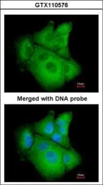
PSAT1 antibody [N1C3]GTX110576
ApplicationsImmunoFluorescence, Western Blot, ImmunoCytoChemistry, ImmunoHistoChemistry, ImmunoHistoChemistry Paraffin
ReactivityHuman, Mouse
TargetPSAT1
- SizePrice
Product group Antibodies
PSAT1 antibody [N3C3]GTX115909
ApplicationsImmunoFluorescence, Western Blot, ImmunoCytoChemistry, ImmunoHistoChemistry, ImmunoHistoChemistry Paraffin
ReactivityHuman, Mouse
TargetPSAT1
- SizePrice