

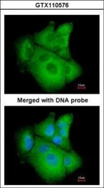
Immunofluorescence analysis of paraformaldehyde-fixed A549, using PSAT1(GTX110576) antibody at 1:200 dilution.
 antibody at 1:500 dilution.
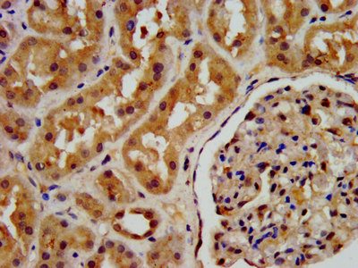
Antigen Retrieval: Trilogy? (EDTA based, pH 8.0) buffer, 15min")
![Non-transfected (–) and transfected (+) 293T whole cell extracts (30 μg) were separated by 10% SDS-PAGE, and the membrane was blotted with PSAT1 antibody [N1C3] (GTX110576) diluted at 1:1000. The HRP-conjugated anti-rabbit IgG antibody (GTX213110-01) was used to detect the primary antibody.](https://www.genetex.com/upload/website/prouct_img/normal/GTX110576/GTX110576_42242_20161124_WB_shRNA_watermark_w_23060500_748.webp "Non-transfected (–) and transfected (+) 293T whole cell extracts (30 μg) were separated by 10% SDS-PAGE, and the membrane was blotted with PSAT1 antibody [N1C3] (GTX110576) diluted at 1:1000. The HRP-conjugated anti-rabbit IgG antibody (GTX213110-01) was used to detect the primary antibody.")
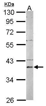
 A: Mouse brain 10% SDS PAGE GTX110576 diluted at 1:1000 The HRP-conjugated anti-rabbit IgG antibody (GTX213110-01) was used to detect the primary antibody.")
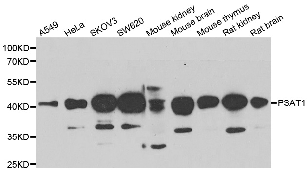
![PSAT1 antibody [N1C3] detects PSAT1 protein by Western blot analysis. Various whole cell extracts (30 μg) were separated by 10% SDS-PAGE, and the membrane was blotted with PSAT1 antibody [N1C3] (GTX110576) diluted at 1:1000.](https://www.genetex.com/upload/website/prouct_img/normal/GTX110576/GTX110576_42243_20151008_WB_25061003_237.webp "PSAT1 antibody [N1C3] detects PSAT1 protein by Western blot analysis. Various whole cell extracts (30 μg) were separated by 10% SDS-PAGE, and the membrane was blotted with PSAT1 antibody [N1C3] (GTX110576) diluted at 1:1000.")
Immunofluorescence analysis of paraformaldehyde-fixed A549, using PSAT1(GTX110576) antibody at 1:200 dilution.
PSAT1 antibody [N1C3]
GTX110576
ApplicationsImmunoFluorescence, Western Blot, ImmunoCytoChemistry, ImmunoHistoChemistry, ImmunoHistoChemistry Paraffin
Product group Antibodies
ReactivityHuman, Mouse
TargetPSAT1
Overview
- SupplierGeneTex
- Product NamePSAT1 antibody [N1C3]
- Delivery Days Customer9
- Application Supplier NoteWB: 1:500-1:3000. ICC/IF: 1:100-1:1000. IHC-P: 1:100-1:1000. *Optimal dilutions/concentrations should be determined by the researcher.Not tested in other applications.
- ApplicationsImmunoFluorescence, Western Blot, ImmunoCytoChemistry, ImmunoHistoChemistry, ImmunoHistoChemistry Paraffin
- CertificationResearch Use Only
- ClonalityPolyclonal
- Concentration0.23 mg/ml
- ConjugateUnconjugated
- Gene ID29968
- Target namePSAT1
- Target descriptionphosphoserine aminotransferase 1
- Target synonymsEPIP, NLS2, PSA, PSAT, PSATD, phosphoserine aminotransferase, endometrial progesterone-induced protein, phosphohydroxythreonine aminotransferase, phosphoserine transaminase
- HostRabbit
- IsotypeIgG
- Protein IDQ9Y617
- Protein NamePhosphoserine aminotransferase
- Scientific DescriptionThe protein encoded by this gene is likely a phosphoserine aminotransferase, based on similarity to proteins in mouse, rabbit, and Drosophila. Alternative splicing of this gene results in two transcript variants encoding different isoforms. [provided by RefSeq]
- ReactivityHuman, Mouse
- Storage Instruction-20°C or -80°C,2°C to 8°C
- UNSPSC41116161
Datasheet
Related products
Product group Antibodies
PSAT1 AntibodyCSB-PA018838LA01HU
ApplicationsImmunoFluorescence, ELISA, ImmunoHistoChemistry
ReactivityHuman
TargetPSAT1
- SizePrice

Product group Antibodies
Anti-PSAT1 Antibody Picoband(r)A06277-CARRIER-FREE
ApplicationsFlow Cytometry, ImmunoFluorescence, Western Blot, ELISA, ImmunoCytoChemistry
ReactivityHuman, Mouse, Rat
TargetPSAT1
- SizePrice
Product group Antibodies
Anti-PSAT1 AntibodyA31537
ApplicationsWestern Blot, ImmunoHistoChemistry
ReactivityHuman, Mouse, Rat
- SizePrice
Product group Antibodies

PSAT1 AntibodyLS-C749133
ApplicationsWestern Blot, ImmunoHistoChemistry
ReactivityHuman, Mouse, Rat
TargetPSAT1
- SizePrice

Product group Antibodies
Anti-PSAT1 AntibodyHPA042924
ApplicationsWestern Blot, ImmunoCytoChemistry
ReactivityHuman
TargetPSAT1
- SizePrice
Product group Antibodies
ApplicationsWestern Blot, ImmunoHistoChemistry
ReactivityMouse, Porcine, Rat
TargetPSAT1
- SizePrice
![Various whole cell extracts (30 μg) were separated by 10% SDS-PAGE, and the membrane was blotted with PSAT1 antibody [HL2270] (GTX638322) diluted at 1:1000. The HRP-conjugated anti-rabbit IgG antibody (GTX213110-01) was used to detect the primary antibody.](https://www.genetex.com/upload/website/prouct_img/normal/GTX638322/GTX638322_T-44970_20230317_WB_23032022_458.webp)
Product group Antibodies
PSAT1 antibody [HL2270]GTX638322
ApplicationsWestern Blot, ImmunoHistoChemistry, ImmunoHistoChemistry Paraffin
ReactivityHuman, Mouse, Rat
TargetPSAT1
- SizePrice
![Various tissue extracts (50 μg) were separated by 10% SDS-PAGE, and the membrane was blotted with PSAT1 antibody [HL2663] (GTX639327) diluted at 1:1000. The HRP-conjugated anti-rabbit IgG antibody (GTX213110-01) was used to detect the primary antibody.](https://www.genetex.com/upload/website/prouct_img/normal/GTX639327/GTX639327_T-45201_20231027_WB_M_R_23103019_286.webp)
Product group Antibodies
PSAT1 antibody [HL2663]GTX639327
ApplicationsImmunoFluorescence, Western Blot, ImmunoCytoChemistry, ImmunoHistoChemistry, ImmunoHistoChemistry Paraffin
ReactivityHuman, Mouse, Rat
TargetPSAT1
- SizePrice
![IHC-P analysis of human cerebrum (grey matter) tissue using GTX640563 PSAT1 antibody [HMV331] HistoMAX?. Strong nuclear and cytoplasmic PSAT1 staining and mainly of astrocytes.](https://www.genetex.com/upload/website/prouct_img/normal/GTX640563/GTX640563_20240703_IHC-P_2_24070300_237.webp)
Product group Antibodies
PSAT1 antibody [HMV331] HistoMAX(tm)GTX640563
ApplicationsImmunoHistoChemistry, ImmunoHistoChemistry Paraffin
ReactivityHuman
TargetPSAT1
- SizePrice
Product group Antibodies
PSAT1 antibody [N3C3]GTX115909
ApplicationsImmunoFluorescence, Western Blot, ImmunoCytoChemistry, ImmunoHistoChemistry, ImmunoHistoChemistry Paraffin
ReactivityHuman, Mouse
TargetPSAT1
- SizePrice