

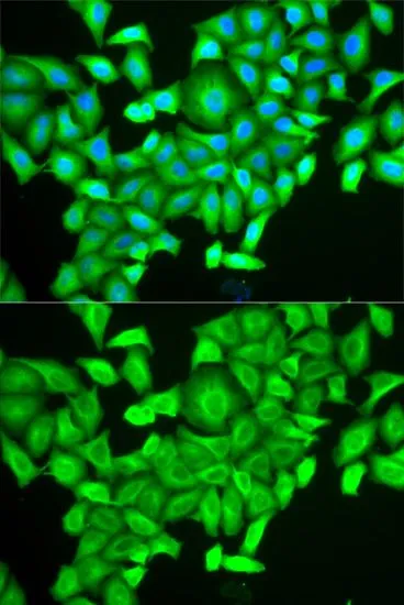
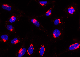
ICC/IF analysis of HeLa cells using GTX32829 RAB6A antibody. Blue : DAPI

ICC/IF analysis of HeLa cells using GTX32829 RAB6A antibody. Blue : DAPI
RAB6A antibody
GTX32829
ApplicationsImmunoFluorescence, Western Blot, ImmunoCytoChemistry
Product group Antibodies
ReactivityHuman, Mouse
TargetRAB6A
Overview
- SupplierGeneTex
- Product NameRAB6A antibody
- Delivery Days Customer9
- Application Supplier NoteWB: 1:500 - 1:2000. ICC/IF: 1:50 - 1:200. *Optimal dilutions/concentrations should be determined by the researcher.Not tested in other applications.
- ApplicationsImmunoFluorescence, Western Blot, ImmunoCytoChemistry
- CertificationResearch Use Only
- ClonalityPolyclonal
- ConjugateUnconjugated
- Gene ID5870
- Target nameRAB6A
- Target descriptionRAB6A, member RAS oncogene family
- Target synonymsRAB6, ras-related protein Rab-6A, RAB6, member RAS oncogene family, Rab GTPase
- HostRabbit
- IsotypeIgG
- Protein IDP20340
- Protein NameRas-related protein Rab-6A
- Scientific DescriptionThis gene encodes a member of the RAB family, which belongs to the small GTPase superfamily. GTPases of the RAB family bind to various effectors to regulate the targeting and fusion of transport carriers to acceptor compartments. This protein is located at the Golgi apparatus, which regulates trafficking in both a retrograde (from early endosomes and Golgi to the endoplasmic reticulum) and an anterograde (from the Golgi to the plasma membrane) directions. Myosin II is an effector of this protein in these processes. This protein is also involved in assembly of human cytomegalovirus (HCMV) by interacting with the cellular protein Bicaudal D1, which interacts with the HCMV virion tegument protein, pp150. Multiple alternatively spliced transcript variants encoding different isoforms have been identified. [provided by RefSeq, Aug 2011]
- ReactivityHuman, Mouse
- Storage Instruction-20°C or -80°C,2°C to 8°C
- UNSPSC41116161
Datasheet
Related products
Product group Antibodies
Anti-RAB6A AntibodyA97306
ApplicationsWestern Blot, ELISA
ReactivityHuman, Mouse, Rat
- SizePrice
Product group Antibodies
anti-Rab6-GTP, mAb (rec.) (AA2)AG-27B-0004
ApplicationsImmunoCytoChemistry
ReactivityDrosophila, Human, Mouse
TargetRAB6A
- SizePrice
Product group Antibodies
Anti-RAB6A Antibody Picoband(r)A02911-1-CARRIER-FREE
ApplicationsWestern Blot, ImmunoHistoChemistry
ReactivityHuman, Mouse, Rat
TargetRAB6A
- SizePrice
Product group Antibodies
RAB6A Polyclonal AntibodyBS-24347R
ApplicationsWestern Blot
ReactivityBovine, Canine, Equine, Human, Mouse, Porcine, Rabbit, Rat, Sheep
TargetRAB6A
- SizePrice
Product group Antibodies
RAB6A AntibodyCSB-PA019216HA01HU
ApplicationsELISA, ImmunoHistoChemistry
ReactivityHuman
TargetRAB6A
- SizePrice
Product group Antibodies

RAB6A / RAB6 AntibodyLS-C402133
ApplicationsWestern Blot, ELISA, ImmunoHistoChemistry
ReactivityHuman, Mouse, Rat
TargetRAB6A
- SizePrice
Product group Antibodies
Anti-RAB6A-25ulHPA059131
ApplicationsWestern Blot, ImmunoCytoChemistry, ImmunoHistoChemistry
ReactivityHuman
- SizePrice
Product group Antibodies
RAB6A antibodyGTX110646
ApplicationsImmunoPrecipitation, Western Blot, ImmunoHistoChemistry, ImmunoHistoChemistry Paraffin
ReactivityHuman, Mouse, Rat
TargetRAB6A
- SizePrice
![Whole zebrafish extract (30 μg) was separated by 12% SDS-PAGE, and the membrane was blotted with RAB6A antibody [HL1047] (GTX635981) diluted at 1:1000. The HRP-conjugated anti-rabbit IgG antibody (GTX213110-01) was used to detect the primary antibody, and the signal was developed with Trident ECL plus-Enhanced.](https://www.genetex.com/upload/website/prouct_img/normal/GTX635981/GTX635981_44256_20221216_WB_Z_22122018_271.webp)
Product group Antibodies
RAB6A antibody [HL1047]GTX635981
ApplicationsWestern Blot, ImmunoHistoChemistry, ImmunoHistoChemistry Paraffin
ReactivityDrosophila, Human, Mouse, Rat, Zebra Fish
TargetRAB6A
- SizePrice