

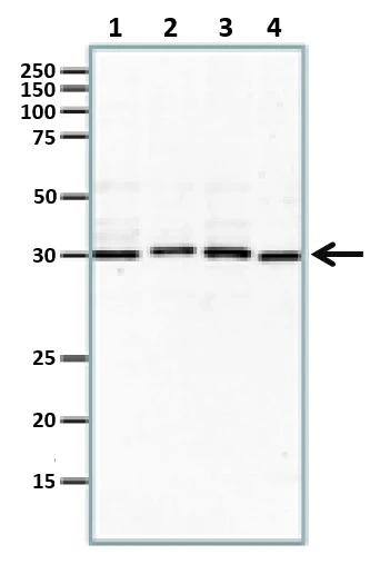
Various tissue extracts (30 μg) were separated by 10% SDS-PAGE, and the membrane was blotted with Rad51 antibody [N1C2] (GTX100469) diluted at 1:1000. The HRP-conjugated anti-rabbit IgG antibody (GTX213110-01) was used to detect the primary antibody, and the signal was developed with Trident ECL plus-Enhanced.
![Rad51 antibody [N1C2] immunoprecipitates Rad51 protein in IP experiments. IP samples: Jurkat whole cell extract A. 40 μg Jurkat whole cell extract B. Control with 4 μg of preimmune Rabbit IgG C. Immunoprecipitation of Rad51 protein by 4 μg Rad51 antibody [N1C2] (GTX100469) 5 % SDS-PAGE The immunoprecipitated Rad51 protein was detected by Rad51 antibody [N1C2] (GTX100469) diluted at 1:500. [EasyBlot anti-rabbit IgG (GTX221666-01) was used as a secondary reagent]](https://www.genetex.com/upload/website/prouct_img/normal/GTX100469/GTX100469_40086_IP_w_23060100_683.webp "Rad51 antibody [N1C2] immunoprecipitates Rad51 protein in IP experiments. IP samples: Jurkat whole cell extract A. 40 μg Jurkat whole cell extract B. Control with 4 μg of preimmune Rabbit IgG C. Immunoprecipitation of Rad51 protein by 4 μg Rad51 antibody [N1C2] (GTX100469) 5 % SDS-PAGE The immunoprecipitated Rad51 protein was detected by Rad51 antibody [N1C2] (GTX100469) diluted at 1:500. [EasyBlot anti-rabbit IgG (GTX221666-01) was used as a secondary reagent]")
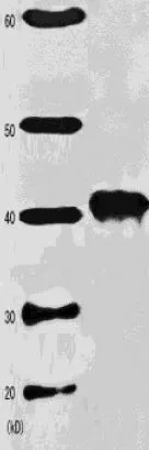
![Various whole cell extracts (30 μg) were separated by 10% SDS-PAGE, and the membrane was blotted with Rad51 antibody [14B4] (GTX100469) diluted at 1:500. The HRP-conjugated anti-rabbit IgG antibody (GTX213110-01) was used to detect the primary antibody.](https://www.genetex.com/upload/website/prouct_img/normal/GTX100469/GTX100469_40086_20170330_WB_M_w_23060100_608.webp "Various whole cell extracts (30 μg) were separated by 10% SDS-PAGE, and the membrane was blotted with Rad51 antibody [14B4] (GTX100469) diluted at 1:500. The HRP-conjugated anti-rabbit IgG antibody (GTX213110-01) was used to detect the primary antibody.")
![Rad51 antibody [N1C2] detects Rad51 protein at nucleus by immunohistochemical analysis. Sample: Paraffin-embedded mouse testis. Rad51 stained by Rad51 antibody [N1C2] (GTX100469) diluted at 1:1000. Antigen Retrieval: Citrate buffer, pH 6.0, 15 min](https://www.genetex.com/upload/website/prouct_img/normal/GTX100469/GTX100469_43467_20190906_IHC-P_M_w_23060100_411.webp "Rad51 antibody [N1C2] detects Rad51 protein at nucleus by immunohistochemical analysis. Sample: Paraffin-embedded mouse testis. Rad51 stained by Rad51 antibody [N1C2] (GTX100469) diluted at 1:1000. Antigen Retrieval: Citrate buffer, pH 6.0, 15 min")
![Various whole cell extracts (30 μg) were separated by 10% SDS-PAGE, and the membrane was blotted with Rad51 antibody [N1C2] (GTX100469) diluted at 1:1000.](https://www.genetex.com/upload/website/prouct_img/normal/GTX100469/GTX100469_42711_20170105_WB_w_23060100_985.webp "Various whole cell extracts (30 μg) were separated by 10% SDS-PAGE, and the membrane was blotted with Rad51 antibody [N1C2] (GTX100469) diluted at 1:1000.")
![Untreated (–) and treated (+) HeLa whole cell extracts (30 μg) were separated by 10% SDS-PAGE, and the membrane was blotted with Rad51 antibody [N1C2] (GTX100469) diluted at 1:1000. The HRP-conjugated anti-rabbit IgG antibody (GTX213110-01) was used to detect the primary antibody.](https://www.genetex.com/upload/website/prouct_img/normal/GTX100469/GTX100469_43516_20190823_WB_treatment_Etoposide_w_23060100_454.webp "Untreated (–) and treated (+) HeLa whole cell extracts (30 μg) were separated by 10% SDS-PAGE, and the membrane was blotted with Rad51 antibody [N1C2] (GTX100469) diluted at 1:1000. The HRP-conjugated anti-rabbit IgG antibody (GTX213110-01) was used to detect the primary antibody.")
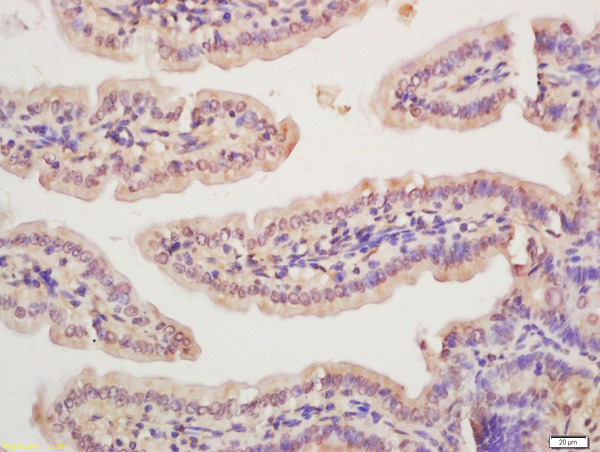
![Rad51 antibody [N1C2] detects Rad51 protein at cytoplasm and nucleus by immunohistochemical analysis. Sample: Paraffin-embedded human cervical carcinoma. Rad51 stained by Rad51 antibody [N1C2] (GTX100469) diluted at 1:500. Antigen Retrieval: Citrate buffer, pH 6.0, 15 min](https://www.genetex.com/upload/website/prouct_img/normal/GTX100469/GTX100469_43467_20190906_IHC-P_w_23060100_219.webp "Rad51 antibody [N1C2] detects Rad51 protein at cytoplasm and nucleus by immunohistochemical analysis. Sample: Paraffin-embedded human cervical carcinoma. Rad51 stained by Rad51 antibody [N1C2] (GTX100469) diluted at 1:500. Antigen Retrieval: Citrate buffer, pH 6.0, 15 min")
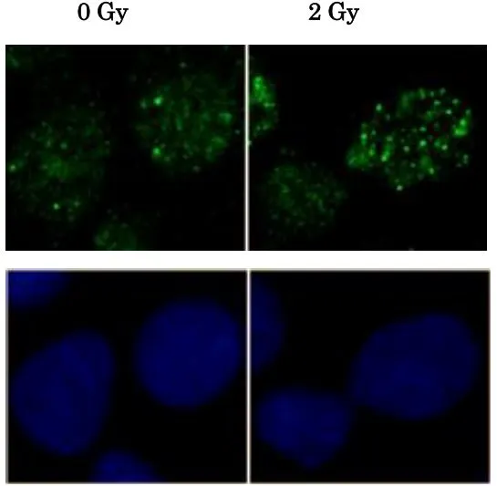
![Rad51 antibody [N1C2] detects Rad51 protein at nucleus by immunofluorescent analysis. Sample: HeLa cells were fixed in 4% paraformaldehyde at RT for 15 min. Green: Rad51 protein stained by Rad51 antibody [N1C2] (GTX100469) diluted at 1:500. Red: phalloidin, a cytoskeleton marker, diluted at 1:200. Scale bar = 10 μm.](https://www.genetex.com/upload/website/prouct_img/normal/GTX100469/GTX100469_40086_20160120_IFA_w_23060100_907.webp "Rad51 antibody [N1C2] detects Rad51 protein at nucleus by immunofluorescent analysis. Sample: HeLa cells were fixed in 4% paraformaldehyde at RT for 15 min. Green: Rad51 protein stained by Rad51 antibody [N1C2] (GTX100469) diluted at 1:500. Red: phalloidin, a cytoskeleton marker, diluted at 1:200. Scale bar = 10 μm.")
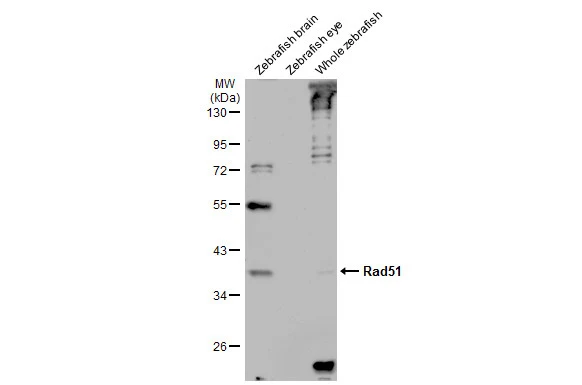
![Various tissue extracts (30 μg) were separated by 10% SDS-PAGE, and the membrane was blotted with Rad51 antibody [N1C2] (GTX100469) diluted at 1:500. The HRP-conjugated anti-rabbit IgG antibody (GTX213110-01) was used to detect the primary antibody, and the signal was developed with Trident ECL plus-Enhanced.](https://www.genetex.com/upload/website/prouct_img/normal/GTX100469/GTX100469_42711_20181026_WB_Z_w_23060100_774.webp "Various tissue extracts (30 μg) were separated by 10% SDS-PAGE, and the membrane was blotted with Rad51 antibody [N1C2] (GTX100469) diluted at 1:500. The HRP-conjugated anti-rabbit IgG antibody (GTX213110-01) was used to detect the primary antibody, and the signal was developed with Trident ECL plus-Enhanced.")
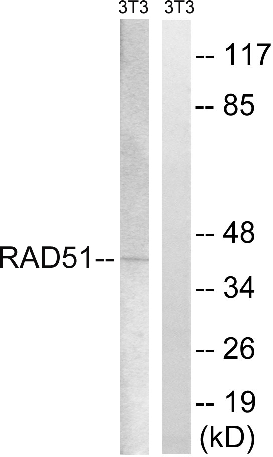
![Various whole cell extracts (30 μg) were separated by 10% SDS-PAGE, and the membrane was blotted with Rad51 antibody [14B4] (GTX100469) diluted at 1:500. The HRP-conjugated anti-rabbit IgG antibody (GTX213110-01) was used to detect the primary antibody.](https://www.genetex.com/upload/website/prouct_img/normal/GTX100469/GTX100469_40086_20170330_WB_R_w_23060100_909.webp "Various whole cell extracts (30 μg) were separated by 10% SDS-PAGE, and the membrane was blotted with Rad51 antibody [14B4] (GTX100469) diluted at 1:500. The HRP-conjugated anti-rabbit IgG antibody (GTX213110-01) was used to detect the primary antibody.")
Various tissue extracts (30 μg) were separated by 10% SDS-PAGE, and the membrane was blotted with Rad51 antibody [N1C2] (GTX100469) diluted at 1:1000. The HRP-conjugated anti-rabbit IgG antibody (GTX213110-01) was used to detect the primary antibody, and the signal was developed with Trident ECL plus-Enhanced.
Rad51 antibody [N1C2]
GTX100469
ApplicationsImmunoFluorescence, ImmunoPrecipitation, Western Blot, ImmunoCytoChemistry, ImmunoHistoChemistry, ImmunoHistoChemistry Paraffin
Product group Antibodies
ReactivityHuman, Mouse, Rat, Zebra Fish
TargetRAD51
Overview
- SupplierGeneTex
- Product NameRad51 antibody [N1C2]
- Delivery Days Customer9
- Application Supplier NoteWB: 1:500-1:3000. ICC/IF: 1:100-1:1000. IHC-P: 1:100-1:1000. IP: 1:100-1:500. *Optimal dilutions/concentrations should be determined by the researcher.Not tested in other applications.
- ApplicationsImmunoFluorescence, ImmunoPrecipitation, Western Blot, ImmunoCytoChemistry, ImmunoHistoChemistry, ImmunoHistoChemistry Paraffin
- CertificationResearch Use Only
- ClonalityPolyclonal
- Concentration0.14 mg/ml
- ConjugateUnconjugated
- Gene ID5888
- Target nameRAD51
- Target descriptionRAD51 recombinase
- Target synonymsBRCC5, FANCR, HRAD51, HsRad51, HsT16930, MRMV2, RAD51A, RECA, DNA repair protein RAD51 homolog 1, BRCA1/BRCA2-containing complex, subunit 5, RAD51 homolog A, RecA, E. coli, homolog of, RecA-like protein, recombination protein A
- HostRabbit
- IsotypeIgG
- Protein IDQ06609
- Protein NameDNA repair protein RAD51 homolog 1
- Scientific DescriptionThe protein encoded by this gene is a member of the RAD51 protein family. RAD51 family members are highly similar to bacterial RecA and Saccharomyces cerevisiae Rad51, and are known to be involved in the homologous recombination and repair of DNA. This protein can interact with the ssDNA-binding protein RPA and RAD52, and it is thought to play roles in homologous pairing and strand transfer of DNA. This protein is also found to interact with BRCA1 and BRCA2, which may be important for the cellular response to DNA damage. BRCA2 is shown to regulate both the intracellular localization and DNA-binding ability of this protein. Loss of these controls following BRCA2 inactivation may be a key event leading to genomic instability and tumorigenesis. Two alternatively spliced transcript variants of this gene, which encode distinct proteins, have been reported. Transcript variants utilizing alternative polyA signals exist. [provided by RefSeq]
- ReactivityHuman, Mouse, Rat, Zebra Fish
- Storage Instruction-20°C or -80°C,2°C to 8°C
- UNSPSC41116161
Datasheet
Related products
Product group Antibodies
Anti-RAD51 AntibodyA98997
ApplicationsWestern Blot, ELISA
ReactivityHuman, Mouse
- SizePrice
Product group Antibodies
Anti-RAD51 Antibody144-06268
ApplicationsImmunoFluorescence, Western Blot
ReactivityHuman, Mouse, Rat
TargetRAD51
- SizePrice
Product group Antibodies

Anti-Rad51 [3C10]AB01312-1.1-BT
ApplicationsImmunoFluorescence, ImmunoPrecipitation, Western Blot, ImmunoHistoChemistry
ReactivityBovine, Human, Porcine
TargetRAD51
- SizePrice

Product group Antibodies
Anti-Rad51 Antibody Picoband(r)A00088-CARRIER-FREE
ApplicationsFlow Cytometry, ImmunoFluorescence, Western Blot, ImmunoCytoChemistry, ImmunoHistoChemistry
ReactivityHuman, Mouse, Rat
TargetRAD51
- SizePrice
Product group Antibodies
Rad51 Polyclonal AntibodyBS-10145R
ApplicationsFlow Cytometry, ImmunoFluorescence, ImmunoCytoChemistry, ImmunoHistoChemistry, ImmunoHistoChemistry Frozen, ImmunoHistoChemistry Paraffin
ReactivityBovine, Canine, Equine, Human, Mouse, Porcine, Rabbit, Rat, Sheep
TargetRAD51
- SizePrice
Product group Antibodies
RAD51 AntibodyCSB-PA003916
ApplicationsWestern Blot, ELISA, ImmunoHistoChemistry
ReactivityHuman, Mouse
TargetRAD51
- SizePrice
Product group Antibodies
RAD51 Polyclonal AntibodyCAC14547
ApplicationsImmunoFluorescence, ImmunoPrecipitation, Western Blot, ELISA, ImmunoHistoChemistry
ReactivityMouse
TargetRAD51
- SizePrice
Product group Antibodies
Rad51 antibodyGTX00719
ApplicationsImmunoFluorescence, ImmunoPrecipitation, Western Blot, ImmunoCytoChemistry
ReactivityChicken, Human, Rodent, Xenopus
TargetRAD51
- SizePrice
Product group Antibodies
Rad51 antibodyGTX00720
ApplicationsDot Blot, ImmunoFluorescence, ImmunoPrecipitation, Western Blot, ChIP Chromatin ImmunoPrecipitation, ELISA, ImmunoCytoChemistry, ImmunoHistoChemistry, ImmunoHistoChemistry Paraffin
ReactivityChicken, Hamster, Human, Mouse, Rodent, Rat, Xenopus
TargetRAD51
- SizePrice
Product group Antibodies
References
Rad51 antibodyGTX00721
ApplicationsImmunoFluorescence, ImmunoPrecipitation, Western Blot, ImmunoCytoChemistry
ReactivityFish, Hamster, Human, Mouse, Rat
TargetRAD51
- SizePrice