

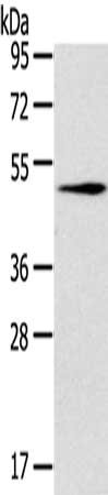
RCC1 antibody detects RCC1 protein by Western blot analysis. A. 30 μg Neuro2A whole cell lysate/extract B. 30 μg GL261 whole cell lysate/extract 12 % SDS-PAGE RCC1 antibody (GTX104590) dilution: 1:1000
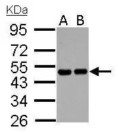
 A: 293T B: A431 (GTX27909) C: H1299 D: HeLa S3 (GTX14654) E: Hep G2 (GTX27900) F: MOLT4 (GTX27912) G: Raji (GTX27908) 10% SDS PAGE GTX104590 diluted at 1:1000")
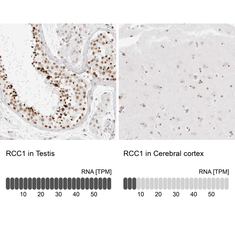
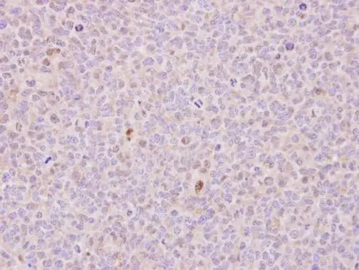
 antibody at 1:500 dilution.
Antigen Retrieval: Trilogy? (EDTA based, pH 8.0) buffer, 15min")
![RCC1 antibody detects RCC1 protein at nucleus by immunofluorescent analysis. Sample: A431 cells were fixed in 4% paraformaldehyde at RT for 15 min. Green: RCC1 protein stained by RCC1 antibody (GTX104590) diluted at 1:500. Red: alpha Tubulin, a cytoskeleton marker, stained by alpha Tubulin antibody [GT114] (GTX628802) diluted at 1:1000.](https://www.genetex.com/upload/website/prouct_img/normal/GTX104590/GTX104590_40002_20160817_IFA_w_23060120_736.webp "RCC1 antibody detects RCC1 protein at nucleus by immunofluorescent analysis. Sample: A431 cells were fixed in 4% paraformaldehyde at RT for 15 min. Green: RCC1 protein stained by RCC1 antibody (GTX104590) diluted at 1:500. Red: alpha Tubulin, a cytoskeleton marker, stained by alpha Tubulin antibody [GT114] (GTX628802) diluted at 1:1000.")
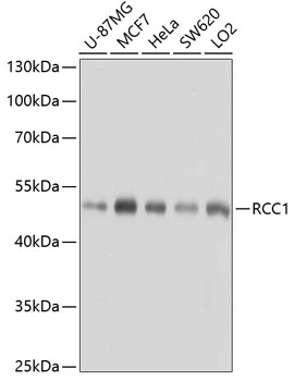
RCC1 antibody detects RCC1 protein by Western blot analysis. A. 30 μg Neuro2A whole cell lysate/extract B. 30 μg GL261 whole cell lysate/extract 12 % SDS-PAGE RCC1 antibody (GTX104590) dilution: 1:1000
RCC1 antibody
GTX104590
ApplicationsImmunoFluorescence, Western Blot, ImmunoCytoChemistry, ImmunoHistoChemistry, ImmunoHistoChemistry Paraffin
Product group Antibodies
ReactivityHuman, Mouse
TargetRCC1
Overview
- SupplierGeneTex
- Product NameRCC1 antibody
- Delivery Days Customer9
- Application Supplier NoteWB: 1:500-1:3000. ICC/IF: 1:100-1:1000. IHC-P: 1:100-1:1000. *Optimal dilutions/concentrations should be determined by the researcher.Not tested in other applications.
- ApplicationsImmunoFluorescence, Western Blot, ImmunoCytoChemistry, ImmunoHistoChemistry, ImmunoHistoChemistry Paraffin
- CertificationResearch Use Only
- ClonalityPolyclonal
- Concentration1 mg/ml
- ConjugateUnconjugated
- Gene ID1104
- Target nameRCC1
- Target descriptionregulator of chromosome condensation 1
- Target synonymsCHC1, IIAAN, RCC1-I, regulator of chromosome condensation, cell cycle regulatory protein, guanine nucleotide-releasing protein
- HostRabbit
- IsotypeIgG
- Protein IDP18754
- Protein NameRegulator of chromosome condensation
- ReactivityHuman, Mouse
- Storage Instruction-20°C or -80°C,2°C to 8°C
- UNSPSC41116161
Datasheet
Related products

Product group Antibodies
Anti-RCC1 AntibodyA38601
ApplicationsWestern Blot, ImmunoHistoChemistry
ReactivityHuman
- SizePrice

Product group Antibodies
Anti-RCC1 Antibody Picoband(r)A02719-1-CARRIER-FREE
ApplicationsFlow Cytometry, ImmunoFluorescence, Western Blot, ELISA, ImmunoCytoChemistry, ImmunoHistoChemistry, ImmunoHistoChemistry Frozen
ReactivityHuman, Mouse, Rat
TargetRCC1
- SizePrice
Product group Antibodies
Anti-RCC1 Antibody144-06350
ApplicationsWestern Blot, ImmunoHistoChemistry
ReactivityHuman, Mouse, Rat
TargetRCC1
- SizePrice
Product group Antibodies

RCC1 AntibodyLS-C813753
ApplicationsWestern Blot, ELISA
ReactivityHuman, Mouse, Rat
TargetRCC1
- SizePrice
Product group Antibodies
RCC1 Recombinant Antibody, AbBy Fluor-647 ConjugatedBSM-62230R-BF647
ApplicationsImmunoFluorescence, Western Blot
ReactivityHuman, Mouse, Rat
TargetRCC1
- SizePrice
Product group Antibodies
RCC1 AntibodyCSB-PA075110
ApplicationsWestern Blot, ELISA
ReactivityHuman, Mouse
TargetRCC1
- SizePrice
Product group Antibodies
Anti-RCC1-25ulHPA027573
ApplicationsWestern Blot, ImmunoCytoChemistry, ImmunoHistoChemistry
ReactivityHuman
- SizePrice
Product group Antibodies
RCC1 antibody [N1C1]GTX113402
ApplicationsWestern Blot, ImmunoHistoChemistry, ImmunoHistoChemistry Paraffin
ReactivityHuman
TargetRCC1
- SizePrice
Product group Antibodies
RCC1 antibodyGTX64560
ApplicationsImmunoFluorescence, Western Blot, ImmunoCytoChemistry, ImmunoHistoChemistry, ImmunoHistoChemistry Paraffin
ReactivityHuman, Mouse, Rat
TargetRCC1
- SizePrice