

Replication Protein A2 (RPA2)(RPA2/2106), 1mg/mL [26628-22-8]
BNUM2106
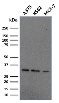
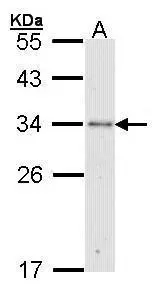
ApplicationsWestern Blot
Product group Antibodies
ReactivityHuman, Mouse
TargetRPA2
Overview
- SupplierBiotium
- Product NameReplication Protein A2 (RPA2)(RPA2/2106), 1mg/mL [26628-22-8]
- Delivery Days Customer9
- ApplicationsWestern Blot
- CertificationResearch Use Only
- ClonalityMonoclonal
- Clone IDRPA2/2106
- Concentration1 mg/ml
- Gene ID6118
- Target nameRPA2
- Target descriptionreplication protein A2
- Target synonymsREPA2, RP-A p32, RP-A p34, RPA32, replication protein A 32 kDa subunit, RF-A protein 2, replication factor A protein 2, replication protein A 34 kDa subunit
- HostMouse
- IsotypeIgG2b
- Protein IDP15927
- Protein NameReplication protein A 32 kDa subunit
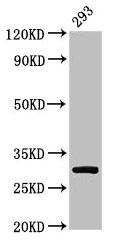
- Scientific DescriptionRecognizes a protein of 34 kDa, identified as a subunit of Replication Protein A (RPA) (also known as human single-stranded DNA binding protein, or HSSB). RPA from human cells is a stable heterotrimer of 70 kDa, 32-34 kDa, and 11-14 kDa subunits (RPA70, RPA32, and RPA14 respectively). It is involved in DNA replication, repair, and recombination. RPA is required for the SV40 large tumor antigen-catalyzed unwinding of SV40 DNA and stimulates DNA polymerase (pol) alpha and delta. RPA34 is phosphorylated at the G1/S boundary of the cell cycle or upon exposure of cells to DNA damage-inducing agents including ionizing and UV radiation. Primary antibodies are available purified, or with a selection of fluorescent CF® Dyes and other labels. CF® Dyes offer exceptional brightness and photostability. Note: Conjugates of blue fluorescent dyes like CF®405S and CF®405M are not recommended for detecting low abundance targets, because blue dyes have lower fluorescence and can give higher non-specific background than other dye colors.
- SourceAnimal
- ReactivityHuman, Mouse
- Storage Instruction-20°C
- UNSPSC12352203
Related products
Product group Antibodies
Anti-RFA2 Antibody102-20182
ApplicationsWestern Blot, ImmunoHistoChemistry, ImmunoHistoChemistry Paraffin
TargetRPA2
- SizePrice

Product group Antibodies
Anti-RPA32/RPA2 Antibody Picoband(r)A02067-1-CARRIER-FREE
ApplicationsFlow Cytometry, ImmunoFluorescence, Western Blot, ELISA, ImmunoCytoChemistry, ImmunoHistoChemistry
ReactivityHuman
TargetRPA2
- SizePrice
Product group Antibodies
References
RPA32 antibody [N1C3]GTX101765
ApplicationsImmunoFluorescence, Western Blot, ImmunoCytoChemistry, ImmunoHistoChemistry, ImmunoHistoChemistry Paraffin
ReactivityChicken, Human
TargetRPA2
- SizePrice
Product group Antibodies
RPA2 Polyclonal AntibodyCAC13840
ApplicationsImmunoFluorescence, ImmunoPrecipitation, Western Blot, ELISA, ImmunoHistoChemistry
TargetRPA2
- SizePrice
Product group Antibodies
Anti-RFA2 AntibodyA96134
ApplicationsImmunoFluorescence, Western Blot, ELISA, ImmunoHistoChemistry
ReactivityHuman, Mouse
- SizePrice
Product group Antibodies
RPA2 Polyclonal AntibodyBS-4182R
ApplicationsImmunoFluorescence, Western Blot, ELISA, ImmunoCytoChemistry, ImmunoHistoChemistry, ImmunoHistoChemistry Frozen, ImmunoHistoChemistry Paraffin
ReactivityBovine, Canine, Chicken, Equine, Human, Mouse, Porcine, Rabbit, Rat
TargetRPA2
- SizePrice
Product group Antibodies
RPA2 AntibodyCSB-PA01555A0RB
ApplicationsImmunoFluorescence, ImmunoPrecipitation, Western Blot, ELISA, ImmunoHistoChemistry
ReactivityHuman
TargetRPA2
- SizePrice
Product group Antibodies

RPA2 / RFA2 / RPA34 AntibodyLS-C770317
ApplicationsWestern Blot, ELISA, ImmunoHistoChemistry
ReactivityHuman
TargetRPA2
- SizePrice