

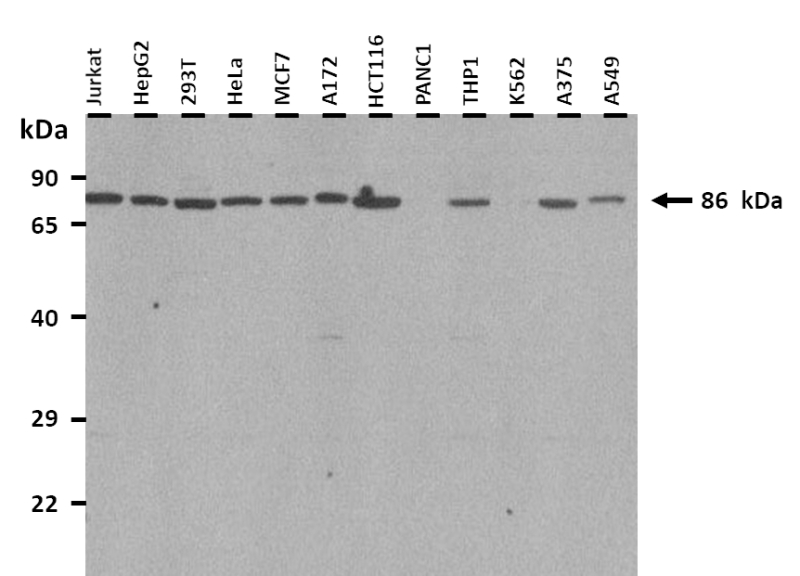
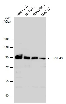
WB analysis of various sample lysates using GTX46918 RNF43 antibody, Internal. Dilution : 1μg/ml Loading : 25μg
WB analysis of various sample lysates using GTX46918 RNF43 antibody, Internal. Dilution : 1μg/ml Loading : 25μg
RNF43 antibody, Internal
GTX46918
ApplicationsWestern Blot
Product group Antibodies
ReactivityHuman
TargetRNF43
Overview
- SupplierGeneTex
- Product NameRNF43 antibody, Internal
- Delivery Days Customer9
- Application Supplier NoteWB: 0.2-2.5 ug/ml. *Optimal dilutions/concentrations should be determined by the researcher.Not tested in other applications.
- ApplicationsWestern Blot
- CertificationResearch Use Only
- ClonalityPolyclonal
- Concentration0.5-1 mg/ml
- ConjugateUnconjugated
- Gene ID54894
- Target nameRNF43
- Target descriptionring finger protein 43
- Target synonymsRNF124, SSPCS, URCC, E3 ubiquitin-protein ligase RNF43, RING-type E3 ubiquitin transferase RNF43
- HostRabbit
- IsotypeIgG
- Protein IDQ68DV7
- Protein NameE3 ubiquitin-protein ligase RNF43
- Scientific DescriptionThe protein encoded by this gene is a RING-type E3 ubiquitin ligase and is predicted to contain a transmembrane domain, a protease-associated domain, an ectodomain, and a cytoplasmic RING domain. This protein is thought to negatively regulate Wnt signaling, and expression of this gene results in an increase in ubiquitination of frizzled receptors, an alteration in their subcellular distribution, resulting in reduced surface levels of these receptors. Mutations in this gene have been reported in multiple tumor cells, including colorectal and endometrial cancers. Alternative splicing results in multiple transcript variants encoding different isoforms. [provided by RefSeq, Mar 2015]
- ReactivityHuman
- Storage Instruction-20°C or -80°C,2°C to 8°C
- UNSPSC41116161
Datasheet
Related products
Product group Antibodies
RNF43 AntibodyCSB-PA019892LA01HU
ApplicationsELISA, ImmunoHistoChemistry
ReactivityHuman
TargetRNF43
- SizePrice

Product group Antibodies
Anti-RNF43 Antibody Picoband(r)A01694-1-CARRIER-FREE
ApplicationsWestern Blot, ELISA
ReactivityHuman
TargetRNF43
- SizePrice

Product group Antibodies
Anti-RNF43 AntibodyHPA008079
ApplicationsWestern Blot, ImmunoHistoChemistry
ReactivityHuman
TargetRNF43
- SizePrice
Product group Antibodies

RNF43 AntibodyLS-C672876
ApplicationsELISA, ImmunoHistoChemistry, ImmunoHistoChemistry Paraffin
ReactivityHuman
TargetRNF43
- SizePrice
Product group Antibodies
RNF43 Polyclonal AntibodyCAC13189
ApplicationsELISA, ImmunoHistoChemistry
TargetRNF43
- SizePrice
Product group Antibodies
RNF43 Polyclonal AntibodyBS-7007R
ApplicationsImmunoFluorescence, Western Blot, ELISA, ImmunoCytoChemistry, ImmunoHistoChemistry, ImmunoHistoChemistry Frozen, ImmunoHistoChemistry Paraffin
ReactivityBovine, Canine, Equine, Human, Mouse, Porcine, Rat
TargetRNF43
- SizePrice
Product group Antibodies
RNF43 antibodyGTX132671
ApplicationsWestern Blot
ReactivityHuman, Mouse
TargetRNF43
- SizePrice
Product group Antibodies
Anti-RNF43 (C-term) Antibody102-21400
ApplicationsWestern Blot, ImmunoHistoChemistry, ImmunoHistoChemistry Paraffin
TargetRNF43
- SizePrice