

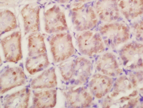
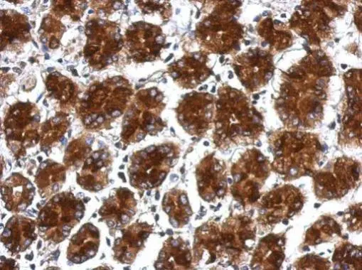
Formalin-fixed and paraffin embedded mouse intestine labeled with Anti-RPLP0 Polyclonal Antibody, Unconjugated (bs-1464R) at 1:200 followed by conjugation to the secondary antibody and DAB staining\n
 at 1:200 followed by conjugation to the secondary antibody and DAB staining\n")
Formalin-fixed and paraffin embedded mouse intestine labeled with Anti-RPLP0 Polyclonal Antibody, Unconjugated (bs-1464R) at 1:200 followed by conjugation to the secondary antibody and DAB staining\n
RPLP0 Polyclonal Antibody
BS-1886R
ApplicationsImmunoFluorescence, Western Blot, ELISA, ImmunoCytoChemistry, ImmunoHistoChemistry, ImmunoHistoChemistry Frozen, ImmunoHistoChemistry Paraffin
Product group Antibodies
ReactivityBovine, Canine, Chicken, Equine, Human, Mouse, Porcine, Rabbit, Rat
TargetRPLP0
Overview
- SupplierBioss
- Product NameRPLP0 Polyclonal Antibody
- Delivery Days Customer16
- ApplicationsImmunoFluorescence, Western Blot, ELISA, ImmunoCytoChemistry, ImmunoHistoChemistry, ImmunoHistoChemistry Frozen, ImmunoHistoChemistry Paraffin
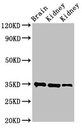
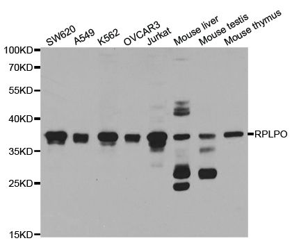
- Applications SupplierWB(1:300-5000), ELISA(1:500-1000), IHC-P(1:200-400), IHC-F(1:100-500), IF(IHC-P)(1:50-200), IF(IHC-F)(1:50-200), IF(ICC)(1:50-200)
- CertificationResearch Use Only
- ClonalityPolyclonal
- Concentration1 ug/ul
- ConjugateUnconjugated
- Gene ID6175
- Target nameRPLP0
- Target descriptionribosomal protein lateral stalk subunit P0
- Target synonymsL10E, LP0, P0, PRLP0, RPP0, uL10, large ribosomal subunit protein uL10, 60S acidic ribosomal protein P0, 60S ribosomal protein L10E, acidic ribosomal phosphoprotein P0, neutral ribosomal phosphoprotein P0, ribosomal protein, large, P0
- HostRabbit
- IsotypeIgG
- Protein IDP05388
- Protein NameLarge ribosomal subunit protein uL10
- ReactivityBovine, Canine, Chicken, Equine, Human, Mouse, Porcine, Rabbit, Rat
- Storage Instruction-20°C
- UNSPSC41116161
Datasheet
Related products
Product group Antibodies
RPLP0 AntibodyCSB-PA020336LA01HU
ApplicationsImmunoFluorescence, Western Blot, ELISA, ImmunoHistoChemistry
ReactivityHuman, Mouse, Rat
TargetRPLP0
- SizePrice
Product group Antibodies
RPLP0 Polyclonal AntibodyCAC13912
ApplicationsImmunoFluorescence, Western Blot, ELISA, ImmunoHistoChemistry
ReactivityMouse, Rat
TargetRPLP0
- SizePrice
Product group Antibodies
Anti-RPLP0 Antibody144-05557
ApplicationsImmunoFluorescence, Western Blot, ImmunoHistoChemistry
ReactivityHuman, Mouse, Rat
TargetRPLP0
- SizePrice
Product group Antibodies
Anti-RPLP0 AntibodyA30818
ApplicationsImmunoFluorescence, Western Blot, ImmunoHistoChemistry
ReactivityHuman, Mouse, Rat
- SizePrice

Product group Antibodies
Anti-RPLP0 AntibodyHPA003512
ApplicationsImmunoCytoChemistry, ImmunoHistoChemistry
ReactivityHuman
TargetRPLP0
- SizePrice

Product group Antibodies
Anti-RPLP0 Antibody Picoband(r)A04349-1-CARRIER-FREE
ApplicationsFlow Cytometry, ImmunoFluorescence, Western Blot, ELISA, ImmunoCytoChemistry
ReactivityHuman
TargetRPLP0
- SizePrice
Product group Antibodies

RPLP0 AntibodyLS-C401015
ApplicationsWestern Blot, ELISA, ImmunoHistoChemistry
ReactivityHuman, Mouse, Rat
TargetRPLP0
- SizePrice
Product group Antibodies
RPLP0 antibodyGTX114730
ApplicationsWestern Blot, ImmunoHistoChemistry, ImmunoHistoChemistry Paraffin
ReactivityHuman
TargetRPLP0
- SizePrice