

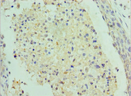
Immunohistochemistry of paraffin-embedded human cervical cancer using CSB-PA020695ESR2HU at dilution of 1:100
Immunohistochemistry of paraffin-embedded human cervical cancer using CSB-PA020695ESR2HU at dilution of 1:100
SAP18 Antibody
CSB-PA020695ESR2HU
ApplicationsELISA, ImmunoHistoChemistry
Product group Antibodies
ReactivityHuman
TargetSAP18
Overview
- SupplierCusabio
- Product NameSAP18 Antibody
- Delivery Days Customer20
- ApplicationsELISA, ImmunoHistoChemistry
- CertificationResearch Use Only
- ClonalityPolyclonal
- ConjugateUnconjugated
- Gene ID10284
- Target nameSAP18
- Target descriptionSin3A associated protein 18
- Target synonyms2HOR0202, SAP18P, histone deacetylase complex subunit SAP18, 18 kDa Sin3-associated polypeptide, Sin3A-associated protein, 18kDa, cell growth inhibiting protein 38, cell growth-inhibiting gene 38 protein, epididymis secretory sperm binding protein, histone deacetlyase complex subunit SAP18, sin3-associated polypeptide, 18 kDa, sin3-associated polypeptide, p18
- HostRabbit
- IsotypeIgG
- Protein IDO00422
- Protein NameHistone deacetylase complex subunit SAP18
- Scientific DescriptionComponent of the SIN3-repressing complex. Enhances the ability of SIN3-HDAC1-mediated transcriptional repression. When tethered to the promoter, it can direct the formation of a repressive complex to core histone proteins. Auxiliary component of the splicing-dependent multiprotein exon junction complex (EJC) deposited at splice junction on mRNAs. The EJC is a dynamic structure consisting of core proteins and several peripheral nuclear and cytoplasmic associated factors that join the complex only transiently either during EJC assembly or during subsequent mRNA metabolism. Component of the ASAP and PSAP complexes which bind RNA in a sequence-independent manner and are proposed to be recruited to the EJC prior to or during the splicing process and to regulate specific excision of introns in specific transcription subsets. The ASAP complex can inhibit mRNA processing during in vitro splicing reactions. The ASAP complex promotes apoptosis and is disassembled after induction of apoptosis. Involved in the splicing modulation of BCL2L1/Bcl-X (and probably other apoptotic genes); specifically inhibits the formation of proapoptotic isoforms such as Bcl-X(S); the activity is different from the established EJC assembly and function.
- ReactivityHuman
- Storage Instruction-20°C or -80°C
- UNSPSC41116161
Related products

Product group Antibodies
Anti-SAP18 Antibody Picoband(r)A06381-1-CARRIER-FREE
ApplicationsFlow Cytometry, ImmunoFluorescence, Western Blot, ELISA, ImmunoCytoChemistry, ImmunoHistoChemistry
ReactivityHuman, Mouse, Rat
TargetSAP18
- SizePrice
Product group Antibodies
Anti-SAP18 Antibody144-65802
ApplicationsImmunoFluorescence, Western Blot, ImmunoHistoChemistry
ReactivityHuman, Mouse, Rat
TargetSAP18
- SizePrice
Product group Antibodies
Anti-SAP18 AntibodyA14557
ApplicationsImmunoFluorescence, Western Blot, ImmunoCytoChemistry, ImmunoHistoChemistry
ReactivityHuman, Mouse, Rat
- SizePrice
Product group Antibodies

SAP18 AntibodyLS-C668278
ApplicationsWestern Blot, ImmunoHistoChemistry, ImmunoHistoChemistry Paraffin
ReactivityHuman
TargetSAP18
- SizePrice
Product group Antibodies
SAP18 Polyclonal AntibodyCAC13193
ApplicationsELISA, ImmunoHistoChemistry
TargetSAP18
- SizePrice

Product group Antibodies
Anti-SAP18 AntibodyHPA011352
ApplicationsImmunoCytoChemistry, ImmunoHistoChemistry
ReactivityHuman
TargetSAP18
- SizePrice
Product group Antibodies
SAP18 antibodyGTX130522
ApplicationsWestern Blot
ReactivityHuman
TargetSAP18
- SizePrice