

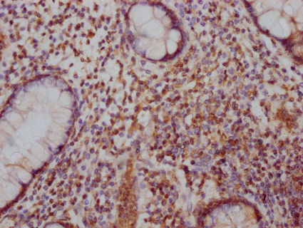
IHC image of CSB-PA892170LA01HU diluted at 1:100 and staining in paraffin-embedded human colon cancer performed on a Leica BondTM system. After dewaxing and hydration, antigen retrieval was mediated by high pressure in a citrate buffer (pH 6.0). Section was blocked with 10% normal goat serum 30min at RT. Then primary antibody (1% BSA) was incubated at 4°C overnight. The primary is detected by a Goat anti-rabbit IgG labeled by HRP and visualized using 0.05% DAB.
. Section was blocked with 10% normal goat serum 30min at RT. Then primary antibody (1% BSA) was incubated at 4°C overnight. The primary is detected by a Goat anti-rabbit IgG labeled by HRP and visualized using 0.05% DAB.")
IHC image of CSB-PA892170LA01HU diluted at 1:100 and staining in paraffin-embedded human colon cancer performed on a Leica BondTM system. After dewaxing and hydration, antigen retrieval was mediated by high pressure in a citrate buffer (pH 6.0). Section was blocked with 10% normal goat serum 30min at RT. Then primary antibody (1% BSA) was incubated at 4°C overnight. The primary is detected by a Goat anti-rabbit IgG labeled by HRP and visualized using 0.05% DAB.
SATB2 Antibody
CSB-PA892170LA01HU
ApplicationsELISA, ImmunoHistoChemistry
Product group Antibodies
ReactivityHuman
TargetSATB2
Overview
- SupplierCusabio
- Product NameSATB2 Antibody
- Delivery Days Customer20
- ApplicationsELISA, ImmunoHistoChemistry
- CertificationResearch Use Only
- ClonalityPolyclonal
- ConjugateUnconjugated
- Gene ID23314
- Target nameSATB2
- Target descriptionSATB homeobox 2
- Target synonymsC2DELq32q33, DEL2Q32Q33, GLSS, DNA-binding protein SATB2, SATB family member 2, SATB2 fusion, special AT-rich sequence-binding protein 2
- HostRabbit
- IsotypeIgG
- Protein IDQ9UPW6
- Protein NameDNA-binding protein SATB2
- Scientific DescriptionBinds to DNA, at nuclear matrix- or scaffold-associated regions. Thought to recognize the sugar-phosphate structure of double-stranded DNA. Transcription factor controlling nuclear gene expression, by binding to matrix attachment regions (MARs) of DNA and inducing a local chromatin-loop remodeling. Acts as a docking site for several chromatin remodeling enzymes and also by recruiting corepressors (HDACs) or coactivators (HATs) directly to promoters and enhancers. Required for the initiation of the upper-layer neurons (UL1) specific genetic program and for the inactivation of deep-layer neurons (DL) and UL2 specific genes, probably by modulating BCL11B expression. Repressor of Ctip2 and regulatory determinant of corticocortical connections in the developing cerebral cortex. May play an important role in palate formation. Acts as a molecular node in a transcriptional network regulating skeletal development and osteoblast differentiation.
- ReactivityHuman
- Storage Instruction-20°C or -80°C
- UNSPSC41116161
Related products
Product group Antibodies
Anti-SATB2 AntibodyA91312
ApplicationsWestern Blot, ImmunoHistoChemistry
ReactivityHuman, Rat
- SizePrice

Product group Antibodies
Anti-SATB2 Antibody Picoband(r)A02588-2-CARRIER-FREE
ApplicationsWestern Blot
ReactivityHuman
TargetSATB2
- SizePrice
Product group Antibodies
Anti-SATB2 Antibody144-64612
ApplicationsWestern Blot
ReactivityHuman
TargetSATB2
- SizePrice

Product group Antibodies
Anti-SATB2 AntibodyAMAB90678
ApplicationsWestern Blot, ImmunoHistoChemistry
ReactivityHuman, Rat
TargetSATB2
- SizePrice
Product group Antibodies
References
SATB2 Polyclonal AntibodyBS-11949R
ApplicationsFlow Cytometry, ImmunoFluorescence, Western Blot, ELISA, ImmunoCytoChemistry, ImmunoHistoChemistry, ImmunoHistoChemistry Frozen, ImmunoHistoChemistry Paraffin
ReactivityBovine, Equine, Human, Mouse, Rat, Sheep
TargetSATB2
- SizePrice
Product group Antibodies
ApplicationsImmunoPrecipitation, Western Blot, ImmunoCytoChemistry, ImmunoHistoChemistry
TargetSATB2
- SizePrice

Product group Antibodies
SATB2 antibodyGTX132972
ApplicationsImmunoFluorescence, Western Blot, ImmunoCytoChemistry
ReactivityHuman, Rat
TargetSATB2
- SizePrice
Product group Antibodies

SATB2 Antibody (aa250-300)LS-C288640
ApplicationsImmunoPrecipitation
ReactivityHuman
TargetSATB2
- SizePrice