

Search results: WAC
Product group Antibodies
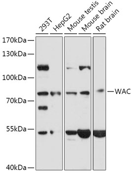
Anti-WAC AntibodyA307981
ApplicationsWestern Blot
ReactivityHuman, Mouse, Rat
- SizePrice

Product group Antibodies
Anti-WAC AntibodyA04941
ApplicationsImmunoFluorescence, Western Blot, ELISA, ImmunoHistoChemistry, ImmunoHistoChemistry Paraffin
ReactivityHuman, Mouse, Rat
TargetWAC
- SizePrice
Product group Antibodies
wac Polyclonal AntibodyCAC13297
ApplicationsWestern Blot, ELISA
ReactivityVirus
Targetwac
- SizePrice
Product group Antibodies
WAC Polyclonal AntibodyBS-12787R
ApplicationsImmunoFluorescence, Western Blot, ELISA, ImmunoCytoChemistry, ImmunoHistoChemistry, ImmunoHistoChemistry Frozen, ImmunoHistoChemistry Paraffin
ReactivityBovine, Canine, Chicken, Equine, Human, Mouse, Porcine, Rabbit, Rat, Sheep
TargetWAC
- SizePrice

Product group Antibodies
Anti-WAC AntibodyHPA036528
ApplicationsImmunoHistoChemistry
ReactivityHuman
TargetWAC
- SizePrice

Product group Antibodies
Anti-WAC AntibodyHPA042609
ApplicationsImmunoCytoChemistry
ReactivityHuman
TargetWAC
- SizePrice
Product group Antibodies
WAC Antibody, HRP conjugatedCSB-PA861133HB01HU
ApplicationsELISA
ReactivityHuman
TargetWAC
- SizePrice
Product group Antibodies
WAC Antibody, FITC conjugatedCSB-PA861133HC01HU
ReactivityHuman
TargetWAC
- SizePrice
Product group Antibodies
WAC Antibody, Biotin conjugatedCSB-PA861133HD01HU
ApplicationsELISA
ReactivityHuman
TargetWAC
- SizePrice

Product group Antibodies
Anti-WAC Antibody Picoband(r)A04941-1-10UG
ApplicationsFlow Cytometry, ImmunoFluorescence, Western Blot, ELISA, ImmunoCytoChemistry
ReactivityHuman, Mouse, Rat
TargetWAC
- SizePrice
Product group Antibodies
Anti-WAC Antibody Picoband(r)A04941-1-APC
ApplicationsFlow Cytometry, ImmunoFluorescence, Western Blot, ELISA, ImmunoCytoChemistry
ReactivityHuman, Mouse, Rat
TargetWAC
- SizePrice
Didn't find what you were looking for?
Search through our product groups to find the right product
Back to overview