

Search results: akt
Product group Antibodies
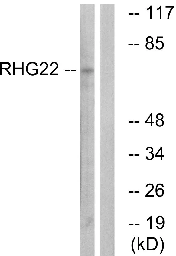
ARHGAP22 antibodyGTX87597
ApplicationsWestern Blot
ReactivityHuman
TargetARHGAP22
- SizePrice
Product group Antibodies
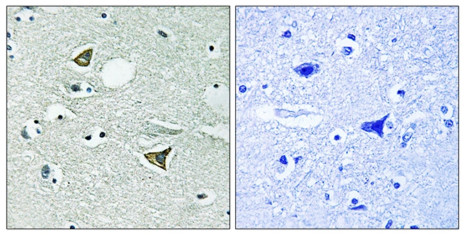
Girdin antibodyGTX87941
ApplicationsImmunoHistoChemistry, ImmunoHistoChemistry Paraffin
ReactivityHuman
TargetCCDC88A
- SizePrice
Product group Antibodies
TCL1A antibody [ZM92]GTX01833
ApplicationsImmunoHistoChemistry, ImmunoHistoChemistry Paraffin
ReactivityHuman
TargetTCL1A
- SizePrice
![WB analysis of MCF-7 whole cell lysate using GTX02584 AKT1 antibody [AKT1/3898R].](https://www.genetex.com/upload/website/prouct_img/normal/GTX02584/GTX02584_20210319_WB_w_23053122_897.webp)
Product group Antibodies
AKT1 antibody [AKT1/3898R]GTX02584
ApplicationsWestern Blot, ELISA
ReactivityHuman
TargetAKT1
- SizePrice
![IHC-P analysis of human breast carcinoma section using GTX02682 MVP/LRP antibody [VP2897R].](https://www.genetex.com/upload/website/prouct_img/normal/GTX02682/GTX02682_20210319_IHC-P_w_23053122_600.webp)
Product group Antibodies
MVP/LRP antibody [VP2897R]GTX02682
ApplicationsImmunoHistoChemistry, ImmunoHistoChemistry Paraffin
ReactivityHuman
TargetMVP
- SizePrice
Product group Antibodies
c-Met antibodyGTX10728
ApplicationsFlow Cytometry, Western Blot, ImmunoHistoChemistry, Neutralisation/Blocking
ReactivityHuman
TargetMET
- SizePrice
Product group Antibodies
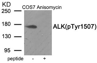
Phospho-ALK (Tyr1507) AntibodyCSB-PA209313
ApplicationsWestern Blot, ELISA
ReactivityHuman, Mouse
TargetALK
- SizePrice
Product group Antibodies
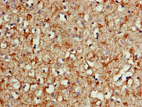
ALK AntibodyCSB-PA21109A0RB
ApplicationsImmunoFluorescence, ELISA, ImmunoHistoChemistry
ReactivityHuman
TargetALK
- SizePrice
Product group Antibodies
ALK Antibody, HRP conjugatedCSB-PA21109B0RB
ApplicationsELISA
ReactivityHuman
TargetALK
- SizePrice
Product group Antibodies
ALK Antibody, FITC conjugatedCSB-PA21109C0RB
ReactivityHuman
TargetALK
- SizePrice
Didn't find what you were looking for?
Search through our product groups to find the right product
Back to overview