

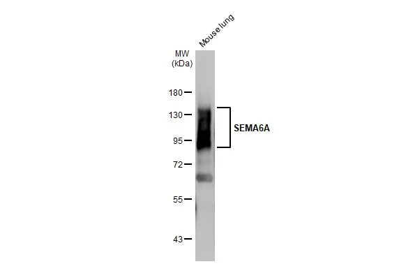
Mouse tissue extract (50 μg) was separated by 7.5% SDS-PAGE, and the membrane was blotted with SEMA6A antibody [HL2120] (GTX638092) diluted at 1:1000. The HRP-conjugated anti-rabbit IgG antibody (GTX213110-01) was used to detect the primary antibody, and the signal was developed with Trident ECL plus-Enhanced.
![Various whole cell extracts (30 μg) were separated by 7.5% SDS-PAGE, and the membrane was blotted with SEMA6A antibody [HL2120] (GTX638092) diluted at 1:1000. The HRP-conjugated anti-rabbit IgG antibody (GTX213110-01) was used to detect the primary antibody. Corresponding RNA expression data for the same cell lines are based on Human Protein Atlas program.](https://www.genetex.com/upload/website/prouct_img/normal/GTX638092/GTX638092_T-44914_20230106_WB_TPM_watermark_23010922_956.webp "Various whole cell extracts (30 μg) were separated by 7.5% SDS-PAGE, and the membrane was blotted with SEMA6A antibody [HL2120] (GTX638092) diluted at 1:1000. The HRP-conjugated anti-rabbit IgG antibody (GTX213110-01) was used to detect the primary antibody. Corresponding RNA expression data for the same cell lines are based on Human Protein Atlas program.")
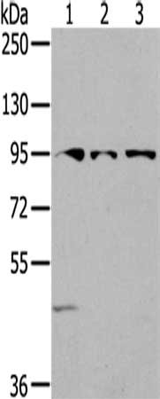
![Whole cell extract (30 μg) was separated by 7.5% SDS-PAGE, and the membrane was blotted with SEMA6A antibody [HL2120] (GTX638092) diluted at 1:1000. The HRP-conjugated anti-rabbit IgG antibody (GTX213110-01) was used to detect the primary antibody, and the signal was developed with Trident ECL plus-Enhanced.](https://www.genetex.com/upload/website/prouct_img/normal/GTX638092/GTX638092_T-44914_20230120_WB_R_23013122_736.webp "Whole cell extract (30 μg) was separated by 7.5% SDS-PAGE, and the membrane was blotted with SEMA6A antibody [HL2120] (GTX638092) diluted at 1:1000. The HRP-conjugated anti-rabbit IgG antibody (GTX213110-01) was used to detect the primary antibody, and the signal was developed with Trident ECL plus-Enhanced.")
Mouse tissue extract (50 μg) was separated by 7.5% SDS-PAGE, and the membrane was blotted with SEMA6A antibody [HL2120] (GTX638092) diluted at 1:1000. The HRP-conjugated anti-rabbit IgG antibody (GTX213110-01) was used to detect the primary antibody, and the signal was developed with Trident ECL plus-Enhanced.
SEMA6A antibody [HL2120]
GTX638092
ApplicationsWestern Blot
Product group Antibodies
ReactivityHuman, Mouse, Rat
TargetSEMA6A
Overview
- SupplierGeneTex
- Product NameSEMA6A antibody [HL2120]
- Delivery Days Customer9
- Application Supplier NoteWB: 1:500-1:3000. *Optimal dilutions/concentrations should be determined by the researcher.Not tested in other applications.
- ApplicationsWestern Blot
- CertificationResearch Use Only
- ClonalityMonoclonal
- Clone IDHL2120
- Concentration1 mg/ml
- ConjugateUnconjugated
- Gene ID57556
- Target nameSEMA6A
- Target descriptionsemaphorin 6A
- Target synonymsHT018, SEMA, SEMA6A1, SEMAQ, VIA, semaphorin-6A, SEMA6A-1, sema VIa, sema domain, transmembrane domain (TM), and cytoplasmic domain, (semaphorin) 6A, semaphorin 6A-1, semaphorin VIA
- HostRabbit
- IsotypeIgG
- Protein IDQ9H2E6
- Protein NameSemaphorin-6A
- Scientific DescriptionThe transmembrane semaphorin SEMA6A is expressed in developing neural tissue and is required for proper development of the thalamocortical projection (Leighton et al., 2001 [PubMed 11242070]).[supplied by OMIM, Feb 2011]
- ReactivityHuman, Mouse, Rat
- Storage Instruction-20°C or -80°C,2°C to 8°C
- UNSPSC41116161
Datasheet
Related products

Product group Antibodies
Anti-Sema6A Antibody Picoband(r)A06967-1-CARRIER-FREE
ApplicationsFlow Cytometry, Western Blot, ELISA, ImmunoHistoChemistry
ReactivityHuman, Mouse, Rat
TargetSEMA6A
- SizePrice
Product group Antibodies
Anti-SEMA6A AntibodyA45603
ApplicationsImmunoHistoChemistry
ReactivityHuman, Mouse
- SizePrice

Product group Antibodies
Anti-SEMA6A AntibodyHPA031265
ApplicationsImmunoHistoChemistry
ReactivityHuman
TargetSEMA6A
- SizePrice
Product group Antibodies
SEMA6A AntibodyCSB-PA049485
ApplicationsWestern Blot, ELISA
ReactivityHuman, Mouse
TargetSEMA6A
- SizePrice
Product group Antibodies

SEMA6A / Semaphorin 6A Antibody (Biotin)LS-C501860
ApplicationsELISA
ReactivityHuman
TargetSEMA6A
- SizePrice
Product group Antibodies
Sema6A Polyclonal AntibodyCAC11058
ApplicationsImmunoFluorescence, ELISA, ImmunoHistoChemistry
TargetSEMA6A
- SizePrice
Product group Antibodies
SEMA6A antibody [8D9]GTX52988
ApplicationsWestern Blot
ReactivityHuman
TargetSEMA6A
- SizePrice
Product group Antibodies
Sema6A Polyclonal AntibodyBS-2701R
ApplicationsImmunoFluorescence, ELISA, ImmunoCytoChemistry, ImmunoHistoChemistry, ImmunoHistoChemistry Frozen, ImmunoHistoChemistry Paraffin
ReactivityBovine, Canine, Human, Mouse, Porcine, Rabbit, Rat
TargetSEMA6A
- SizePrice