

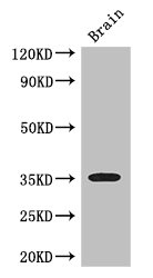
Western Blot Positive WB detected in: Mouse brain tissue All lanes: SFRP1 antibody at 3ug/ml Secondary Goat polyclonal to rabbit IgG at 1/50000 dilution Predicted band size: 36 kDa Observed band size: 36 kDa
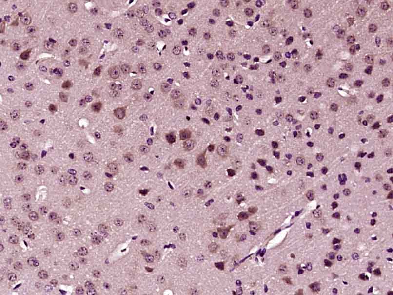
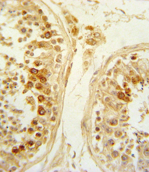
. Section was blocked with 10% normal goat serum 30min at RT. Then primary antibody (1% BSA) was incubated at 4°C overnight. The primary is detected by a biotinylated secondary antibody and visualized using an HRP conjugated SP system.")
")
Western Blot Positive WB detected in: Mouse brain tissue All lanes: SFRP1 antibody at 3ug/ml Secondary Goat polyclonal to rabbit IgG at 1/50000 dilution Predicted band size: 36 kDa Observed band size: 36 kDa
SFRP1 Antibody
CSB-PA021138LA01HU
ApplicationsImmunoFluorescence, Western Blot, ELISA, ImmunoHistoChemistry
Product group Antibodies
ReactivityHuman, Mouse
TargetSFRP1
Overview
- SupplierCusabio
- Product NameSFRP1 Antibody
- Delivery Days Customer20
- ApplicationsImmunoFluorescence, Western Blot, ELISA, ImmunoHistoChemistry
- CertificationResearch Use Only
- ClonalityPolyclonal
- ConjugateUnconjugated
- Gene ID6422
- Target nameSFRP1
- Target descriptionsecreted frizzled related protein 1
- Target synonymsFRP, FRP-1, FRP1, FrzA, SARP2, secreted frizzled-related protein 1, SARP-2, frizzled-related protein, sFRP-1, secreted apoptosis-related protein 2
- HostRabbit
- IsotypeIgG
- Protein IDQ8N474
- Protein NameSecreted frizzled-related protein 1
- Scientific DescriptionSoluble frizzled-related proteins (sFRPS) function as modulators of Wnt signaling through direct interaction with Wnts. They have a role in regulating cell growth and differentiation in specific cell types. SFRP1 decreases intracellular beta-catenin levels (By similarity). Has antiproliferative effects on vascular cells, in vitro and in vivo, and can induce, in vivo, an angiogenic response. In vascular cell cycle, delays the G1 phase and entry into the S phase (By similarity). In kidney development, inhibits tubule formation and bud growth in metanephroi (By similarity). Inhibits WNT1/WNT4-mediated TCF-dependent transcription.
- ReactivityHuman, Mouse
- Storage Instruction-20°C or -80°C
- UNSPSC41116161
Related products

Product group Antibodies
Anti-SFRP1 Antibody Picoband(r)A01968-2-CARRIER-FREE
ApplicationsFlow Cytometry, ImmunoFluorescence, Western Blot, ELISA, ImmunoCytoChemistry, ImmunoHistoChemistry
ReactivityHuman, Mouse, Rat
TargetSFRP1
- SizePrice
Product group Antibodies
SFRP1 Polyclonal AntibodyBS-1303R
ApplicationsImmunoFluorescence, Western Blot, ELISA, ImmunoCytoChemistry, ImmunoHistoChemistry, ImmunoHistoChemistry Frozen, ImmunoHistoChemistry Paraffin
ReactivityHuman, Mouse, Rat
TargetSFRP1
- SizePrice
Product group Antibodies

Goat anti-SFRP1EB08864
ApplicationsELISA, ImmunoHistoChemistry
ReactivityBovine, Human, Mouse
TargetSFRP1
- SizePrice
Product group Antibodies
SFRP1 Polyclonal AntibodyCAC14649
ApplicationsImmunoFluorescence, Western Blot, ELISA, ImmunoHistoChemistry
ReactivityMouse
TargetSFRP1
- SizePrice
Product group Antibodies

SFRP1 AntibodyLS-C406022
ApplicationsELISA, ImmunoHistoChemistry
ReactivityHuman, Mouse
TargetSFRP1
- SizePrice
Product group Antibodies
SFRP1 antibodyGTX03397
ApplicationsFlow Cytometry, Western Blot, ImmunoHistoChemistry, ImmunoHistoChemistry Paraffin
ReactivityHuman
TargetSFRP1
- SizePrice