

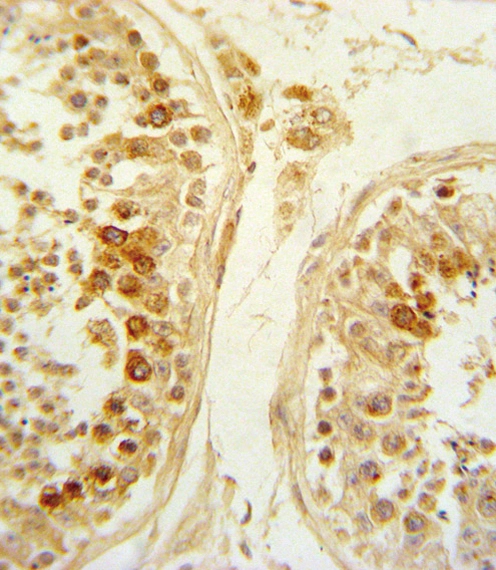
IHC-P analysis of human testis tissue using GTX03397 SFRP1 antibody.
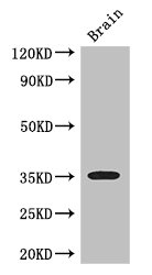
 using GTX03397 SFRP1 antibody.")
 compared to a negative control cell (top histogram) using GTX03397 SFRP1 antibody.")
IHC-P analysis of human testis tissue using GTX03397 SFRP1 antibody.
SFRP1 antibody
GTX03397
ApplicationsFlow Cytometry, Western Blot, ImmunoHistoChemistry, ImmunoHistoChemistry Paraffin
Product group Antibodies
ReactivityHuman
TargetSFRP1
Overview
- SupplierGeneTex
- Product NameSFRP1 antibody
- Delivery Days Customer9
- Application Supplier NoteWB: 1:1000. IHC-P: 1:10-1:50. FCM: 1:10-1:50. *Optimal dilutions/concentrations should be determined by the researcher.Not tested in other applications.
- ApplicationsFlow Cytometry, Western Blot, ImmunoHistoChemistry, ImmunoHistoChemistry Paraffin
- CertificationResearch Use Only
- ClonalityPolyclonal
- ConjugateUnconjugated
- Gene ID6422
- Target nameSFRP1
- Target descriptionsecreted frizzled related protein 1
- Target synonymsFRP, FRP-1, FRP1, FrzA, SARP2, secreted frizzled-related protein 1, SARP-2, frizzled-related protein, sFRP-1, secreted apoptosis-related protein 2
- HostRabbit
- IsotypeIgG
- Protein IDQ8N474
- Protein NameSecreted frizzled-related protein 1
- Scientific DescriptionThis gene encodes a member of the SFRP family that contains a cysteine-rich domain homologous to the putative Wnt-binding site of Frizzled proteins. Members of this family act as soluble modulators of Wnt signaling; epigenetic silencing of SFRP genes leads to deregulated activation of the Wnt-pathway which is associated with cancer. This gene may also be involved in determining the polarity of photoreceptor cells in the retina. [provided by RefSeq, Sep 2009]
- ReactivityHuman
- Storage Instruction-20°C or -80°C,2°C to 8°C
- UNSPSC41116161
Datasheet
Related products
Product group Antibodies
SFRP1 AntibodyCSB-PA021138LA01HU
ApplicationsImmunoFluorescence, Western Blot, ELISA, ImmunoHistoChemistry
ReactivityHuman, Mouse
TargetSFRP1
- SizePrice

Product group Antibodies
Anti-SFRP1 Antibody Picoband(r)A01968-2-CARRIER-FREE
ApplicationsFlow Cytometry, ImmunoFluorescence, Western Blot, ELISA, ImmunoCytoChemistry, ImmunoHistoChemistry
ReactivityHuman, Mouse, Rat
TargetSFRP1
- SizePrice
Product group Antibodies

Goat anti-SFRP1EB08864
ApplicationsELISA, ImmunoHistoChemistry
ReactivityBovine, Human, Mouse
TargetSFRP1
- SizePrice
Product group Antibodies

SFRP1 AntibodyLS-C406022
ApplicationsELISA, ImmunoHistoChemistry
ReactivityHuman, Mouse
TargetSFRP1
- SizePrice
Product group Antibodies
SFRP1 Polyclonal AntibodyBS-1303R
ApplicationsImmunoFluorescence, Western Blot, ELISA, ImmunoCytoChemistry, ImmunoHistoChemistry, ImmunoHistoChemistry Frozen, ImmunoHistoChemistry Paraffin
ReactivityHuman, Mouse, Rat
TargetSFRP1
- SizePrice

Product group Antibodies
References
SFRP1 antibodyGTX24193
ApplicationsImmunoFluorescence, Western Blot, ELISA, ImmunoCytoChemistry, ImmunoHistoChemistry, ImmunoHistoChemistry Paraffin
ReactivityHuman
TargetSFRP1
- SizePrice
Product group Antibodies
SFRP1 Polyclonal AntibodyCAC14649
ApplicationsImmunoFluorescence, Western Blot, ELISA, ImmunoHistoChemistry
ReactivityMouse
TargetSFRP1
- SizePrice