

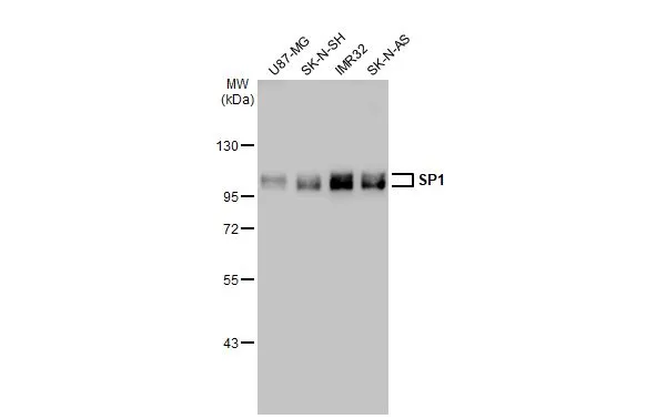
Various whole cell extracts (30 μg) were separated by 7.5% SDS-PAGE, and the membrane was blotted with SP1 antibody (GTX110593) diluted at 1:2000. The HRP-conjugated anti-rabbit IgG antibody (GTX213110-01) was used to detect the primary antibody.
 diluted at 1:500. Antigen Retrieval: Citrate buffer, pH 6.0, 15 min")

![SP1 antibody detects SP1 protein at nucleus by immunofluorescent analysis. Sample: HeLa cells were fixed in 4% paraformaldehyde at RT for 15 min. Green: SP1 stained by SP1 antibody (GTX110593) diluted at 1:100. Red: alpha Tubulin, a cytoskeleton marker, stained by alpha Tubulin antibody [GT114] (GTX628802) diluted at 1:1000. Blue: Fluoroshield with DAPI (GTX30920).](https://www.genetex.com/upload/website/prouct_img/normal/GTX110593/GTX110593_44398_20211126_ICC_IF_w_23060500_876.webp "SP1 antibody detects SP1 protein at nucleus by immunofluorescent analysis. Sample: HeLa cells were fixed in 4% paraformaldehyde at RT for 15 min. Green: SP1 stained by SP1 antibody (GTX110593) diluted at 1:100. Red: alpha Tubulin, a cytoskeleton marker, stained by alpha Tubulin antibody [GT114] (GTX628802) diluted at 1:1000. Blue: Fluoroshield with DAPI (GTX30920).")
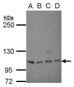
. Western blot analysis was performed using OCT1 antibody (GTX110593) diluted at 1:250. EasyBlot anti-Rabbit IgG (GTX221666-01) was used as a secondary reagent.")
 diluted at 1:1000. Red: phalloidin, a cytoskeleton marker, diluted at 1:200.")
 diluted at 1:2000. Antigen Retrieval: Citrate buffer, pH 6.0, 15 min")
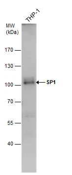
 and transfected (+) 293T whole cell extracts (50 μg) were separated by 7.5% SDS-PAGE, and the membrane was blotted with SP1 antibody (GTX110593) diluted at 1:10000. The HRP-conjugated anti-rabbit IgG antibody (GTX213110-01) was used to detect the primary antibody, and the signal was developed with Trident ECL plus-Enhanced.")
 antibody at 1:500 dilution.
Antigen Retrieval: Trilogy? (EDTA based, pH 8.0) buffer, 15min")

Various whole cell extracts (30 μg) were separated by 7.5% SDS-PAGE, and the membrane was blotted with SP1 antibody (GTX110593) diluted at 1:2000. The HRP-conjugated anti-rabbit IgG antibody (GTX213110-01) was used to detect the primary antibody.
SP1 antibody
GTX110593
ApplicationsImmunoFluorescence, ImmunoPrecipitation, Western Blot, ChIP Chromatin ImmunoPrecipitation, ImmunoCytoChemistry, ImmunoHistoChemistry, ImmunoHistoChemistry Paraffin
Product group Antibodies
ReactivityHuman
TargetSP1
Overview
- SupplierGeneTex
- Product NameSP1 antibody
- Delivery Days Customer9
- Application Supplier NoteWB: 1:500-1:3000. ICC/IF: 1:100-1:1000. IHC-P: 1:100-1:1000. IP: 1:100-1:500. *Optimal dilutions/concentrations should be determined by the researcher.Not tested in other applications.
- ApplicationsImmunoFluorescence, ImmunoPrecipitation, Western Blot, ChIP Chromatin ImmunoPrecipitation, ImmunoCytoChemistry, ImmunoHistoChemistry, ImmunoHistoChemistry Paraffin
- CertificationResearch Use Only
- ClonalityPolyclonal
- Concentration0.22 mg/ml
- ConjugateUnconjugated
- Gene ID6667
- Target nameSP1
- Target descriptionSp1 transcription factor
- Target synonymstranscription factor Sp1, specificity protein 1
- HostRabbit
- IsotypeIgG
- Protein IDP08047
- Protein NameTranscription factor Sp1
- Scientific DescriptionTranscription factor that can activate or repress transcription in response to physiological and pathological stimuli. Binds with high affinity to GC-rich motifs and regulates the expression of a large number of genes involved in a variety of processes such as cell growth, apoptosis, differentiation and immune responses. Highly regulated by post-translational modifications (phosphorylations, sumoylation, proteolytic cleavage, glycosylation and acetylation). Binds also the PDGFR-alpha G-box promoter. May have a role in modulating the cellular response to DNA damage. Implicated in chromatin remodeling. Plays a role in the recruitment of SMARCA4/BRG1 on the c-FOS promoter. Plays an essential role in the regulation of FE65 gene expression.
- ReactivityHuman
- Storage Instruction-20°C or -80°C,2°C to 8°C
- UNSPSC12352203
References
- Wu R, Lin H, Zhang W, et al. Cooperation of long noncoding RNA LOC100909675 and transcriptional regulator CTCF modulates Cdk1 transcript to control astrocyte proliferation. J Biol Chem. 2023,299(9):105153. doi: 10.1016/j.jbc.2023.105153Read this paper
- Moon SJ, Choi HJ, Kye YH, et al. CTTN Overexpression Confers Cancer Stem Cell-like Properties and Trastuzumab Resistance via DKK-1/WNT Signaling in HER2 Positive Breast Cancer. Cancers (Basel). 2023,15(4). doi: 10.3390/cancers15041168Read this paper
- Li Y, Wang Y, Gao L, et al. Betulinic acid self-assembled nanoparticles for effective treatment of glioblastoma. J Nanobiotechnology. 2022,20(1):39. doi: 10.1186/s12951-022-01238-7Read this paper
- González-González L, Gallego-Gutiérrez H, Martin-Tapia D, et al. ZO-2 favors Hippo signaling, and its re-expression in the steatotic liver by AMPK restores junctional sealing. Tissue Barriers. 2022,10(2):1994351. doi: 10.1080/21688370.2021.1994351Read this paper
- Liu L, Deng Y, Zheng Z, et al. Hsp90 Inhibitor STA9090 Sensitizes Hepatocellular Carcinoma to Hyperthermia-Induced DNA Damage by Suppressing DNA-PKcs Protein Stability and mRNA Transcription. Mol Cancer Ther. 2021,20(10):1880-1892. doi: 10.1158/1535-7163.MCT-21-0215Read this paper
- Lee J, Jung E, Gestoso K, et al. ZEB1 Mediates Fibrosis in Corneal Endothelial Mesenchymal Transition Through SP1 and SP3. Invest Ophthalmol Vis Sci. 2020,61(8):41. doi: 10.1167/iovs.61.8.41Read this paper
- Chien SY, Tsai CH, Liu SC, et al. Noggin Inhibits IL-1β and BMP-2 Expression, and Attenuates Cartilage Degeneration and Subchondral Bone Destruction in Experimental Osteoarthritis. Cells. 2020,9(4). doi: 10.3390/cells9040927Read this paper
- Seira N, Yamagata K, Fukushima K, et al. Cellular density-dependent increases in HIF-1α compete with c-Myc to down-regulate human EP4 receptor promoter activity through Sp-1-binding region. Pharmacol Res Perspect. 2018,6(6):e00441. doi: 10.1002/prp2.441Read this paper
- Carrasco-Torres G, Monroy-Ramírez HC, Martínez-Guerra AA, et al. Quercetin Reverses Rat Liver Preneoplastic Lesions Induced by Chemical Carcinogenesis. Oxid Med Cell Longev. 2017,2017:4674918. doi: 10.1155/2017/4674918Read this paper
Datasheet
Related products
Product group Antibodies
Anti-SP1 Antibody144-63395
ApplicationsWestern Blot, ImmunoHistoChemistry
ReactivityHuman, Mouse
TargetSP1
- SizePrice

Product group Antibodies
Anti-SP1 Antibody Picoband(r)A00110-1-CARRIER-FREE
ApplicationsFlow Cytometry, ImmunoFluorescence, Western Blot, ELISA, ImmunoCytoChemistry, ImmunoHistoChemistry
ReactivityHuman, Mouse, Rat
TargetSP1
- SizePrice
![ICC/IF analysis of PFA-fixed MCF-7 cells using GTX01012 SP1 antibody [JF0950]. Green : primary antibody Blue : DAPI Permeabilization : 0.25% Triton X-100 in PBS](https://www.genetex.com/upload/website/prouct_img/normal/GTX01012/GTX01012_20200303_ICC-IF_171_w_23053121_394.webp)
Product group Antibodies
SP1 antibody [JF0950]GTX01012
ApplicationsImmunoFluorescence, Western Blot, ImmunoCytoChemistry, ImmunoHistoChemistry, ImmunoHistoChemistry Paraffin
ReactivityHuman
TargetSP1
- SizePrice
![IHC-P analysis of rat spleen tissue section using GTX01528 SP1 antibody [GT1220]. Dilution : 1:100](https://www.genetex.com/upload/website/prouct_img/normal/GTX01528/GTX01528_20200508_IHC-P_1_w_23053121_526.webp)
Product group Antibodies
SP1 antibody [GT1220]GTX01528
ApplicationsImmunoFluorescence, Western Blot, ImmunoCytoChemistry, ImmunoHistoChemistry, ImmunoHistoChemistry Paraffin
ReactivityHuman, Mouse, Rat
TargetSP1
- SizePrice
Product group Antibodies
SP1 antibody [C1C2], InternalGTX100671
ApplicationsWestern Blot
ReactivityHuman
TargetSP1
- SizePrice
Product group Antibodies
SP1 antibodyGTX103031
ApplicationsImmunoFluorescence, Western Blot, ImmunoCytoChemistry, ImmunoHistoChemistry, ImmunoHistoChemistry Paraffin
ReactivityHuman
TargetSP1
- SizePrice
![SP1 antibody [GT2574] detects SP1 protein at cytoplasm and nucleus by immunohistochemical analysis. Sample: Paraffin-embedded human esophageal carcinoma. SP1 stained by SP1 antibody [GT2574] (GTX634352) diluted at 1:200. Antigen Retrieval: Citrate buffer, pH 6.0, 15 min](https://www.genetex.com/upload/website/prouct_img/normal/GTX634352/GTX634352_42947_20181130_IHC-P_w_23061202_913.webp)
Product group Antibodies
SP1 antibody [GT2574]GTX634352
ApplicationsImmunoFluorescence, Western Blot, ImmunoCytoChemistry, ImmunoHistoChemistry, ImmunoHistoChemistry Paraffin
ReactivityHuman
TargetSP1
- SizePrice
![SP1 antibody [HL1396] detects SP1 protein at nucleus by immunohistochemical analysis. Sample: Paraffin-embedded mouse intestine. SP1 stained by SP1 antibody [HL1396] (GTX636836) diluted at 1:100. Antigen Retrieval: Citrate buffer, pH 6.0, 15 min](https://www.genetex.com/upload/website/prouct_img/normal/GTX636836/GTX636836_44662_20220527_IHC-P_M_22062121_567.webp)
Product group Antibodies
References
SP1 antibody [HL1396]GTX636836
ApplicationsImmunoFluorescence, Western Blot, ImmunoCytoChemistry, ImmunoHistoChemistry, ImmunoHistoChemistry Paraffin
ReactivityHuman, Mouse, Rat
TargetSP1
- SizePrice
Product group Antibodies
Anti-SP1Y058141
ApplicationsWestern Blot, ELISA
ReactivityHuman, Mouse, Rat
- SizePrice