

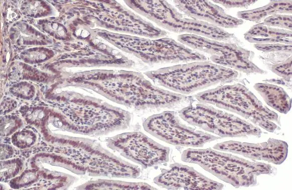
SP1 antibody [HL1396] detects SP1 protein at nucleus by immunohistochemical analysis. Sample: Paraffin-embedded mouse intestine. SP1 stained by SP1 antibody [HL1396] (GTX636836) diluted at 1:100. Antigen Retrieval: Citrate buffer, pH 6.0, 15 min
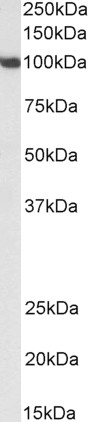
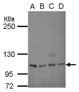
![Various whole cell extracts (30 μg) were separated by 7.5% SDS-PAGE, and the membrane was blotted with SP1 antibody [HL1396] (GTX636836) diluted at 1:10000. The HRP-conjugated anti-rabbit IgG antibody (GTX213110-01) was used to detect the primary antibody.](https://www.genetex.com/upload/website/prouct_img/normal/GTX636836/GTX636836_44748_20220722_WB_22080923_641.webp "Various whole cell extracts (30 μg) were separated by 7.5% SDS-PAGE, and the membrane was blotted with SP1 antibody [HL1396] (GTX636836) diluted at 1:10000. The HRP-conjugated anti-rabbit IgG antibody (GTX213110-01) was used to detect the primary antibody.")
![Non-transfected (–) and transfected (+) 293T whole cell extracts (30 μg) were separated by 7.5% SDS-PAGE, and the membrane was blotted with SP1 antibody [HL1396] (GTX636836) diluted at 1:10000. The HRP-conjugated anti-rabbit IgG antibody (GTX213110-01) was used to detect the primary antibody.](https://www.genetex.com/upload/website/prouct_img/normal/GTX636836/GTX636836_44748_20220819_WB_shRNA_watermark_22082402_182.webp "Non-transfected (–) and transfected (+) 293T whole cell extracts (30 μg) were separated by 7.5% SDS-PAGE, and the membrane was blotted with SP1 antibody [HL1396] (GTX636836) diluted at 1:10000. The HRP-conjugated anti-rabbit IgG antibody (GTX213110-01) was used to detect the primary antibody.")
![SP1 antibody [HL1396] detects SP1 protein at nucleus by immunohistochemical analysis. Sample: Paraffin-embedded human breast carcinoma. SP1 stained by SP1 antibody [HL1396] (GTX636836) diluted at 1:500. Antigen Retrieval: Citrate buffer, pH 6.0, 15 min](https://www.genetex.com/upload/website/prouct_img/normal/GTX636836/GTX636836_44748_20220805_IHC-P_1_22083119_503.webp "SP1 antibody [HL1396] detects SP1 protein at nucleus by immunohistochemical analysis. Sample: Paraffin-embedded human breast carcinoma. SP1 stained by SP1 antibody [HL1396] (GTX636836) diluted at 1:500. Antigen Retrieval: Citrate buffer, pH 6.0, 15 min")
![SP1 antibody [HL1396] detects SP1 protein at nucleus by immunohistochemical analysis. Sample: Paraffin-embedded human breast carcinoma. SP1 stained by SP1 antibody [HL1396] (GTX636836) diluted at 1:500. Antigen Retrieval: Citrate buffer, pH 6.0, 15 min](https://www.genetex.com/upload/website/prouct_img/normal/GTX636836/GTX636836_44748_20220805_IHC-P_22083119_299.webp "SP1 antibody [HL1396] detects SP1 protein at nucleus by immunohistochemical analysis. Sample: Paraffin-embedded human breast carcinoma. SP1 stained by SP1 antibody [HL1396] (GTX636836) diluted at 1:500. Antigen Retrieval: Citrate buffer, pH 6.0, 15 min")
![SP1 antibody [HL1396] detects SP1 protein at nucleus by immunofluorescent analysis. Sample: HeLa cells were fixed in 4% paraformaldehyde at RT for 15 min. Green: SP1 stained by SP1 antibody [HL1396] (GTX636836) diluted at 1:500. Red: alpha Tubulin, a cytoskeleton marker, stained by alpha Tubulin antibody [GT114] (GTX628802) diluted at 1:1000.](https://www.genetex.com/upload/website/prouct_img/normal/GTX636836/GTX636836_44748_20221104_ICC_IF_22112219_116.webp "SP1 antibody [HL1396] detects SP1 protein at nucleus by immunofluorescent analysis. Sample: HeLa cells were fixed in 4% paraformaldehyde at RT for 15 min. Green: SP1 stained by SP1 antibody [HL1396] (GTX636836) diluted at 1:500. Red: alpha Tubulin, a cytoskeleton marker, stained by alpha Tubulin antibody [GT114] (GTX628802) diluted at 1:1000.")
![SP1 antibody [HL1396] detects SP1 protein by immunohistochemical analysis. Sample: Paraffin-embedded rat tissue. SP1 stained by SP1 antibody [HL1396] (GTX636836) diluted at 1:100. Antigen Retrieval: Citrate buffer, pH 6.0, 15 min](https://www.genetex.com/upload/website/prouct_img/normal/GTX636836/GTX636836_44748_20221228_IHC-P_multiple_R_22122901_959.webp "SP1 antibody [HL1396] detects SP1 protein by immunohistochemical analysis. Sample: Paraffin-embedded rat tissue. SP1 stained by SP1 antibody [HL1396] (GTX636836) diluted at 1:100. Antigen Retrieval: Citrate buffer, pH 6.0, 15 min")
![SP1 antibody [HL1396] detects SP1 protein by immunohistochemical analysis. Sample: Paraffin-embedded mouse tissue. SP1 stained by SP1 antibody [HL1396] (GTX636836) diluted at 1:100. Antigen Retrieval: Citrate buffer, pH 6.0, 15 min](https://www.genetex.com/upload/website/prouct_img/normal/GTX636836/GTX636836_44748_20221228_IHC-P_multiple_M_22122901_188.webp "SP1 antibody [HL1396] detects SP1 protein by immunohistochemical analysis. Sample: Paraffin-embedded mouse tissue. SP1 stained by SP1 antibody [HL1396] (GTX636836) diluted at 1:100. Antigen Retrieval: Citrate buffer, pH 6.0, 15 min")
![Non-transfected (–) and transfected (+) 293T whole cell extracts were separated by 7.5% SDS-PAGE, and the membrane was blotted with SP1 antibody [HL1396] (GTX636836) diluted at 1:5000. The HRP-conjugated anti-rabbit IgG antibody (GTX213110-01) was used to detect the primary antibody.](https://www.genetex.com/upload/website/prouct_img/normal/GTX636836/GTX636836_44748_20240119_WB_multiple_B_24012217_881.webp "Non-transfected (–) and transfected (+) 293T whole cell extracts were separated by 7.5% SDS-PAGE, and the membrane was blotted with SP1 antibody [HL1396] (GTX636836) diluted at 1:5000. The HRP-conjugated anti-rabbit IgG antibody (GTX213110-01) was used to detect the primary antibody.")
SP1 antibody [HL1396] detects SP1 protein at nucleus by immunohistochemical analysis. Sample: Paraffin-embedded mouse intestine. SP1 stained by SP1 antibody [HL1396] (GTX636836) diluted at 1:100. Antigen Retrieval: Citrate buffer, pH 6.0, 15 min
SP1 antibody [HL1396]
GTX636836
ApplicationsImmunoFluorescence, Western Blot, ImmunoCytoChemistry, ImmunoHistoChemistry, ImmunoHistoChemistry Paraffin
Product group Antibodies
ReactivityHuman, Mouse, Rat
TargetSP1
Overview
- SupplierGeneTex
- Product NameSP1 antibody [HL1396]
- Delivery Days Customer9
- Application Supplier NoteWB: 1:1000-1:10000. *Optimal dilutions/concentrations should be determined by the researcher.Not tested in other applications.
- ApplicationsImmunoFluorescence, Western Blot, ImmunoCytoChemistry, ImmunoHistoChemistry, ImmunoHistoChemistry Paraffin
- CertificationResearch Use Only
- ClonalityMonoclonal
- Clone IDHL1396
- Concentration0.5 mg/ml
- ConjugateUnconjugated
- Gene ID6667
- Target nameSP1
- Target descriptionSp1 transcription factor
- Target synonymstranscription factor Sp1, specificity protein 1
- HostRabbit
- IsotypeIgG
- Protein IDP08047
- Protein NameTranscription factor Sp1
- Scientific DescriptionThe protein encoded by this gene is a zinc finger transcription factor that binds to GC-rich motifs of many promoters. The encoded protein is involved in many cellular processes, including cell differentiation, cell growth, apoptosis, immune responses, response to DNA damage, and chromatin remodeling. Post-translational modifications such as phosphorylation, acetylation, glycosylation, and proteolytic processing significantly affect the activity of this protein, which can be an activator or a repressor. Three transcript variants encoding different isoforms have been found for this gene. [provided by RefSeq, Nov 2014]
- ReactivityHuman, Mouse, Rat
- Storage Instruction-20°C or -80°C,2°C to 8°C
- UNSPSC41116161
Datasheet
Related products

Product group Antibodies
Anti-SP1 Antibody Picoband(r)A00110-1-CARRIER-FREE
ApplicationsFlow Cytometry, ImmunoFluorescence, Western Blot, ELISA, ImmunoCytoChemistry, ImmunoHistoChemistry
ReactivityHuman, Mouse, Rat
TargetSP1
- SizePrice
Product group Antibodies
Anti-SP1 AntibodyA84617
ApplicationsWestern Blot, ELISA
ReactivityMouse
- SizePrice
Product group Antibodies

Goat anti-SP1EB06834
ApplicationsWestern Blot, ELISA
ReactivityBovine, Canine, Human, Mouse, Porcine, Rat
TargetSP1
- SizePrice

Product group Antibodies
Anti-SP1 AntibodyHPA001853
ApplicationsWestern Blot, ChIP Chromatin ImmunoPrecipitation, ImmunoCytoChemistry, ImmunoHistoChemistry
ReactivityHuman
TargetSP1
- SizePrice
Product group Antibodies
Phospho-SP1 (T453) AntibodyCSB-PA050123
ApplicationsImmunoFluorescence, Western Blot, ELISA, ImmunoHistoChemistry
ReactivityHuman, Mouse, Rat
TargetSP1
- SizePrice
Product group Antibodies

SP1 AntibodyLS-C402792
ApplicationsELISA, ImmunoHistoChemistry
ReactivityHuman, Mouse, Rat
TargetSP1
- SizePrice
Product group Antibodies
SP1 antibodyGTX26082
ApplicationsWestern Blot
ReactivityHuman, Mouse, Rat
TargetSP1
- SizePrice
Product group Antibodies
ApplicationsImmunoPrecipitation, Western Blot, ImmunoCytoChemistry, ImmunoHistoChemistry
ReactivityPorcine
TargetSP1
- SizePrice
Product group Antibodies
SP1 antibody [C1C2], InternalGTX100671
ApplicationsWestern Blot
ReactivityHuman
TargetSP1
- SizePrice