

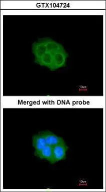
Immunofluorescence analysis of paraformaldehyde-fixed A431, using SPHK1(GTX104724) antibody at 1:200 dilution.
 antibody at 1:100 dilution.
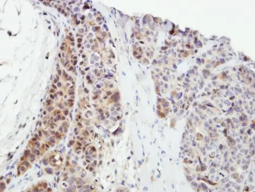
Antigen Retrieval: Trilogy? (EDTA based, pH 8.0) buffer, 15min")
![Non-transfected (–) and transfected (+) HepG2 whole cell extracts (30 μg) were separated by 10% SDS-PAGE, and the membrane was blotted with SPHK1 antibody [C3], C-term (GTX104724) diluted at 1:1000. The HRP-conjugated anti-rabbit IgG antibody (GTX213110-01) was used to detect the primary antibody.](https://www.genetex.com/upload/website/prouct_img/normal/GTX104724/GTX104724_45049_20230609_WB_shRNA_watermark_23061400_585.webp "Non-transfected (–) and transfected (+) HepG2 whole cell extracts (30 μg) were separated by 10% SDS-PAGE, and the membrane was blotted with SPHK1 antibody [C3], C-term (GTX104724) diluted at 1:1000. The HRP-conjugated anti-rabbit IgG antibody (GTX213110-01) was used to detect the primary antibody.")
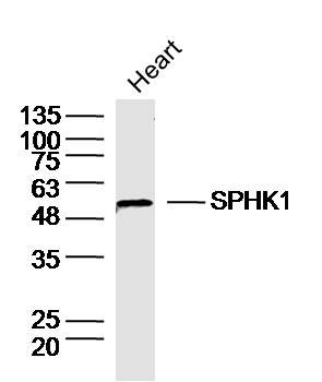
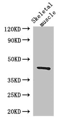
![Various whole cell extracts (30 μg) were separated by 10% SDS-PAGE, and the membrane was blotted with SPHK1 antibody [C3], C-term (GTX104724) diluted at 1:1000. The HRP-conjugated anti-rabbit IgG antibody (GTX213110-01) was used to detect the primary antibody. Corresponding RNA expression data for the same cell lines are based on Human Protein Atlas program.](https://www.genetex.com/upload/website/prouct_img/normal/GTX104724/GTX104724_45091_20230707_WB_TPM_watermark_25042920_317.webp "Various whole cell extracts (30 μg) were separated by 10% SDS-PAGE, and the membrane was blotted with SPHK1 antibody [C3], C-term (GTX104724) diluted at 1:1000. The HRP-conjugated anti-rabbit IgG antibody (GTX213110-01) was used to detect the primary antibody. Corresponding RNA expression data for the same cell lines are based on Human Protein Atlas program.")
Immunofluorescence analysis of paraformaldehyde-fixed A431, using SPHK1(GTX104724) antibody at 1:200 dilution.
SPHK1 antibody [C3], C-term
GTX104724
ApplicationsImmunoFluorescence, Western Blot, ImmunoCytoChemistry, ImmunoHistoChemistry, ImmunoHistoChemistry Paraffin
Product group Antibodies
ReactivityHuman
TargetSPHK1
Overview
- SupplierGeneTex
- Product NameSPHK1 antibody [C3], C-term
- Delivery Days Customer9
- Application Supplier NoteWB: 1:500-1:3000. ICC/IF: 1:100-1:1000. IHC-P: 1:100-1:1000. *Optimal dilutions/concentrations should be determined by the researcher.Not tested in other applications.
- ApplicationsImmunoFluorescence, Western Blot, ImmunoCytoChemistry, ImmunoHistoChemistry, ImmunoHistoChemistry Paraffin
- CertificationResearch Use Only
- ClonalityPolyclonal
- Concentration1.05 mg/ml
- ConjugateUnconjugated
- Gene ID8877
- Target nameSPHK1
- Target descriptionsphingosine kinase 1
- Target synonymsSPHK, sphingosine kinase 1, SK 1, SPK 1, acetyltransferase SPHK1
- HostRabbit
- IsotypeIgG
- Protein IDQ9NYA1
- Protein NameSphingosine kinase 1
- Scientific DescriptionSphingosine-1-phosphate (SPP) is a novel lipid messenger with both intracellular and extracellular functions. Intracellularly, it regulates proliferation and survival, and extracellularly, it is a ligand for EDG1 (MIM 601974). Various stimuli increase cellular levels of SPP by activation of sphingosine kinase (SPHK), the enzyme that catalyzes the phosphorylation of sphingosine. Competitive inhibitors of SPHK block formation of SPP and selectively inhibit cellular proliferation induced by a variety of factors, including platelet-derived growth factor (e.g., MIM 173430) and serum.[supplied by OMIM]
- ReactivityHuman
- Storage Instruction-20°C or -80°C,2°C to 8°C
- UNSPSC41116161
Datasheet
Related products
Product group Antibodies
Anti-SPHK1 AntibodyA83416
ApplicationsImmunoPrecipitation, Western Blot, ELISA
ReactivityHuman
- SizePrice

Product group Antibodies
Anti-SPHK1 Antibody Picoband(r)A01390-1-CARRIER-FREE
ApplicationsFlow Cytometry, ImmunoFluorescence, Western Blot, ELISA, ImmunoCytoChemistry, ImmunoHistoChemistry
ReactivityHuman, Mouse, Rat
TargetSPHK1
- SizePrice
Product group Antibodies
Anti-SPHK1 Antibody144-00139
ApplicationsWestern Blot, ImmunoHistoChemistry
ReactivityHuman, Mouse, Rat
TargetSPHK1
- SizePrice
Product group Antibodies
References
SPHK1 Polyclonal AntibodyBS-2652R
ApplicationsFlow Cytometry, ImmunoFluorescence, Western Blot, ELISA, ImmunoCytoChemistry, ImmunoHistoChemistry, ImmunoHistoChemistry Frozen, ImmunoHistoChemistry Paraffin
ReactivityBovine, Canine, Human, Mouse, Rabbit, Rat
TargetSPHK1
- SizePrice
Product group Antibodies

Goat anti-SPHK1EB08409
ApplicationsImmunoPrecipitation, Western Blot, ELISA
ReactivityBovine, Canine, Human, Mouse, Rat
TargetSPHK1
- SizePrice
Product group Antibodies
SPHK1 Polyclonal AntibodyCAC14602
ApplicationsImmunoFluorescence, Western Blot, ELISA, ImmunoHistoChemistry
ReactivityMouse
TargetSPHK1
- SizePrice
Product group Antibodies
SPHK1 AntibodyCSB-PA022564LA01HU
ApplicationsImmunoFluorescence, Western Blot, ELISA, ImmunoHistoChemistry
ReactivityHuman, Mouse
TargetSPHK1
- SizePrice

Product group Antibodies
SPHK1 antibody, InternalGTX88593
ApplicationsImmunoPrecipitation, Western Blot
ReactivityHuman
TargetSPHK1
- SizePrice
Product group Antibodies
SPHK1 antibodyGTX107509
ApplicationsImmunoFluorescence, Western Blot, ImmunoCytoChemistry, ImmunoHistoChemistry, ImmunoHistoChemistry Paraffin
ReactivityHuman
TargetSPHK1
- SizePrice