

Mouse heart lysates probed with SPHK1 Polyclonal Antibody, unconjugated (bs-2652R) at 1:30 overnight at 4°C followed by a conjugated secondary antibody at 1:10000 for 90 minutes at 37°C.\n
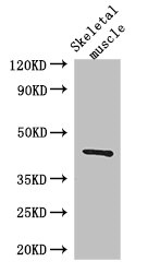
 at 1:1000 dilution and 4°C overnight incubation. Followed by conjugated secondary antibody incubation at 1:20000 for 60 min at 37˚C.")
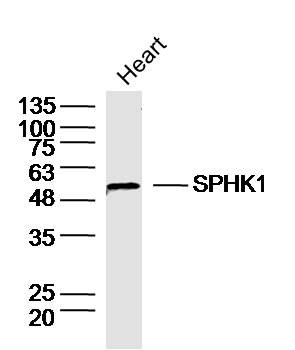
Mouse heart lysates probed with SPHK1 Polyclonal Antibody, unconjugated (bs-2652R) at 1:30 overnight at 4°C followed by a conjugated secondary antibody at 1:10000 for 90 minutes at 37°C.\n
SPHK1 Polyclonal Antibody
BS-2652R
ApplicationsFlow Cytometry, ImmunoFluorescence, Western Blot, ELISA, ImmunoCytoChemistry, ImmunoHistoChemistry, ImmunoHistoChemistry Frozen, ImmunoHistoChemistry Paraffin
Product group Antibodies
ReactivityBovine, Canine, Human, Mouse, Rabbit, Rat
TargetSPHK1
Overview
- SupplierBioss
- Product NameSPHK1 Polyclonal Antibody
- Delivery Days Customer16
- ApplicationsFlow Cytometry, ImmunoFluorescence, Western Blot, ELISA, ImmunoCytoChemistry, ImmunoHistoChemistry, ImmunoHistoChemistry Frozen, ImmunoHistoChemistry Paraffin
- Applications SupplierWB(1:300-5000), ELISA(1:500-1000), FCM(1:20-100), IHC-P(1:200-400), IHC-F(1:100-500), IF(IHC-P)(1:50-200), IF(IHC-F)(1:50-200), IF(ICC)(1:50-200)
- CertificationResearch Use Only
- ClonalityPolyclonal
- Concentration1 ug/ul
- ConjugateUnconjugated
- Gene ID8877
- Target nameSPHK1
- Target descriptionsphingosine kinase 1
- Target synonymsSPHK, sphingosine kinase 1, SK 1, SPK 1, acetyltransferase SPHK1
- HostRabbit
- IsotypeIgG
- Protein IDQ9NYA1
- Protein NameSphingosine kinase 1
- ReactivityBovine, Canine, Human, Mouse, Rabbit, Rat
- Storage Instruction-20°C
- UNSPSC41116161
References
- Disrupted epithelial/macrophage crosstalk via Spinster homologue 2-mediated S1P signaling may drive defective macrophage phagocytic function in COPD. Tran HB et al., 2017, PLoS OneRead this paper
- Hair Cell Loss Induced by Sphingosine and a Sphingosine Kinase Inhibitor in the Rat Cochlea. Tani K et al., 2016 Jan, Neurotox ResRead this paper
Datasheet
Related products
Product group Antibodies
SPHK1 AntibodyCSB-PA022564LA01HU
ApplicationsImmunoFluorescence, Western Blot, ELISA, ImmunoHistoChemistry
ReactivityHuman, Mouse
TargetSPHK1
- SizePrice

Product group Antibodies
Anti-SPHK1 Antibody Picoband(r)A01390-1-CARRIER-FREE
ApplicationsFlow Cytometry, ImmunoFluorescence, Western Blot, ELISA, ImmunoCytoChemistry, ImmunoHistoChemistry
ReactivityHuman, Mouse, Rat
TargetSPHK1
- SizePrice
Product group Antibodies
Anti-SPHK1 Antibody144-00139
ApplicationsWestern Blot, ImmunoHistoChemistry
ReactivityHuman, Mouse, Rat
TargetSPHK1
- SizePrice
Product group Antibodies
Anti-SPHK1 AntibodyA83416
ApplicationsImmunoPrecipitation, Western Blot, ELISA
ReactivityHuman
- SizePrice
Product group Antibodies

SPHK / SPHK1 AntibodyLS-C746745
ApplicationsImmunoFluorescence, Western Blot
ReactivityHuman, Mouse
TargetSPHK1
- SizePrice
Product group Antibodies

Goat anti-SPHK1EB08409
ApplicationsImmunoPrecipitation, Western Blot, ELISA
ReactivityBovine, Canine, Human, Mouse, Rat
TargetSPHK1
- SizePrice
Product group Antibodies
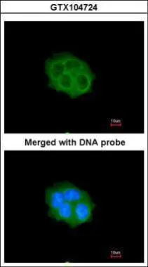
SPHK1 antibody [C3], C-termGTX104724
ApplicationsImmunoFluorescence, Western Blot, ImmunoCytoChemistry, ImmunoHistoChemistry, ImmunoHistoChemistry Paraffin
ReactivityHuman
TargetSPHK1
- SizePrice