

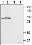
WB analysis of rat brain (lanes 1, 3) and RBL cell (lanes 2, 4) lysates using GTX54768 STIM2 antibody preincubated with or without immunogen peptide. Dilution : 1:200


WB analysis of rat brain (lanes 1, 3) and RBL cell (lanes 2, 4) lysates using GTX54768 STIM2 antibody preincubated with or without immunogen peptide. Dilution : 1:200
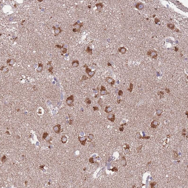
STIM2 antibody
GTX54768
ApplicationsImmunoFluorescence, Western Blot, ImmunoCytoChemistry, ImmunoHistoChemistry, ImmunoHistoChemistry Paraffin
Product group Antibodies
ReactivityHuman, Mouse, Rat
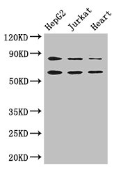
TargetSTIM2
Overview
- SupplierGeneTex
- Product NameSTIM2 antibody
- Delivery Days Customer7
- ApplicationsImmunoFluorescence, Western Blot, ImmunoCytoChemistry, ImmunoHistoChemistry, ImmunoHistoChemistry Paraffin
- CertificationResearch Use Only
- ClonalityPolyclonal
- Concentration0.8 mg/ml
- ConjugateUnconjugated
- Gene ID57620
- Target nameSTIM2
- Target descriptionstromal interaction molecule 2
- Target synonymsstromal interaction molecule 2
- HostRabbit
- IsotypeIgG
- Protein IDQ9P246
- Protein NameStromal interaction molecule 2
- Scientific DescriptionThis gene is a member of the stromal interaction molecule (STIM) family and likely arose, along with related family member STIM1, from a common ancestral gene. The encoded protein functions to regulate calcium concentrations in the cytosol and endoplasmic reticulum, and is involved in the activation of plasma membrane Orai Ca(2+) entry channels. This gene initiates translation from a non-AUG (UUG) start site. A signal peptide is cleaved from the resulting protein. Multiple transcript variants result from alternative splicing. [provided by RefSeq, Dec 2009]
- ReactivityHuman, Mouse, Rat
- Storage Instruction-20°C or -80°C,2°C to 8°C
- UNSPSC41116161
Datasheet
Related products
Product group Antibodies
Anti-STIM2 Antibody144-65131
ApplicationsImmunoFluorescence, Western Blot
ReactivityHuman, Mouse, Rat
TargetSTIM2
- SizePrice

Product group Antibodies
Anti-STIM2 Antibody Picoband(r)A02345-4-CARRIER-FREE
ApplicationsImmunoFluorescence, Western Blot, ELISA, ImmunoCytoChemistry
ReactivityHuman, Rat
TargetSTIM2
- SizePrice
Product group Antibodies
STIM2 Polyclonal AntibodyCAC14651
ApplicationsWestern Blot, ELISA, ImmunoHistoChemistry
ReactivityMouse
TargetSTIM2
- SizePrice
Product group Antibodies
STIM2 AntibodyCSB-PA022830LA01HU
ApplicationsWestern Blot, ELISA, ImmunoHistoChemistry
ReactivityHuman, Mouse
TargetSTIM2
- SizePrice
Product group Antibodies

STIM2 Antibody (Biotin)LS-C499875
ApplicationsELISA
ReactivityHuman
TargetSTIM2
- SizePrice
Product group Antibodies
References
STIM2 antibodyGTX85440
ApplicationsWestern Blot, ELISA, ImmunoHistoChemistry, ImmunoHistoChemistry Paraffin
ReactivityHuman, Mouse, Rat
TargetSTIM2
- SizePrice
Product group Antibodies
STIM2 antibodyGTX85441
ApplicationsWestern Blot, ELISA, ImmunoHistoChemistry, ImmunoHistoChemistry Paraffin
ReactivityHuman, Mouse, Rat
TargetSTIM2
- SizePrice
Product group Antibodies
Anti-STIM2 AntibodyHPA036933
ApplicationsWestern Blot, ImmunoHistoChemistry
ReactivityHuman
TargetSTIM2
- SizePrice