

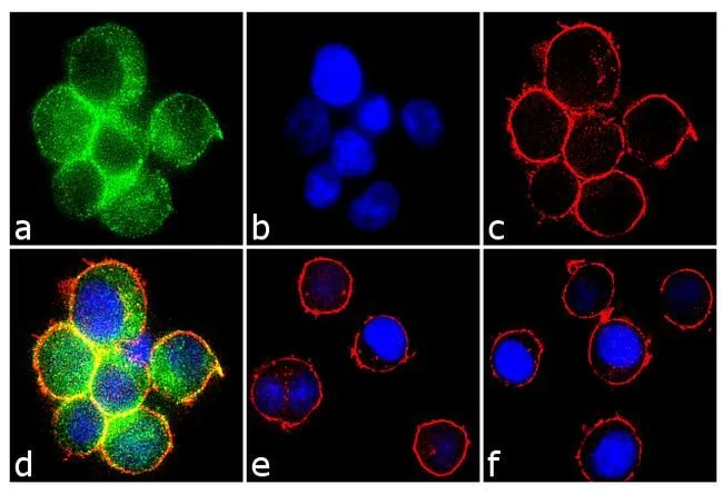
ICC/IF analysis of Jurkat cells treated with 100 uM H2O2 for 1 hour using GTX12862 Syk (phospho Tyr323) antibody. Panel e shows untreated cells with no signal. Panel f represents control cells with no primary antibody to assess background. Green : Primary antibody Blue : Nuclei Red : Actin Fixation : 4% paraformaldehyde Permeabilization : 0.1% Trito X-100 for 10 minutesDilution: 2 μg/ml
 analysis of Jurkat cells treated with 10 mM H2O2 for 3 minutes (Lane 2-6) using GTX12862 Syk (phospho Tyr323) antibody prior incubated with the non-phosphopeptide corresponding to the phosphopeptide immunogen (Lane 3), a generic phosphotyrosine-containing peptide (Lane 4), or the phosphopeptide immunogen (Lane 5) control. The data show that only the immunogen phosphopeptide blocks the signal, demonstrating the specificity of the antibody. The membrane treated with lambda phosphatase (Lane 6) eliminates the signal further verifying that the antibody is phospho-specific.")
ICC/IF analysis of Jurkat cells treated with 100 uM H2O2 for 1 hour using GTX12862 Syk (phospho Tyr323) antibody. Panel e shows untreated cells with no signal. Panel f represents control cells with no primary antibody to assess background. Green : Primary antibody Blue : Nuclei Red : Actin Fixation : 4% paraformaldehyde Permeabilization : 0.1% Trito X-100 for 10 minutesDilution: 2 μg/ml
Syk (phospho Tyr323) antibody
GTX12862
ApplicationsImmunoFluorescence, Western Blot, ImmunoCytoChemistry
Product group Antibodies
ReactivityHuman, Mouse
TargetSYK
Overview
- SupplierGeneTex
- Product NameSyk (phospho Tyr323) antibody
- Delivery Days Customer9
- Application Supplier NoteICC/IF: 1:250. *Optimal dilutions/concentrations should be determined by the researcher.Not tested in other applications.
- ApplicationsImmunoFluorescence, Western Blot, ImmunoCytoChemistry
- CertificationResearch Use Only
- ClonalityPolyclonal
- ConjugateUnconjugated
- Gene ID6850
- Target nameSYK
- Target descriptionspleen associated tyrosine kinase
- Target synonymsIMD82, p72-Syk, tyrosine-protein kinase SYK, spleen tyrosine kinase
- HostRabbit
- IsotypeIgG
- Protein IDP43405
- Protein NameTyrosine-protein kinase SYK
- Scientific DescriptionThis gene encodes a member of the family of non-receptor type Tyr protein kinases. This protein is widely expressed in hematopoietic cells and is involved in coupling activated immunoreceptors to downstream signaling events that mediate diverse cellular responses, including proliferation, differentiation, and phagocytosis. It is thought to be a modulator of epithelial cell growth and a potential tumour suppressor in human breast carcinomas. Alternatively spliced transcript variants encoding different isoforms have been found for this gene. [provided by RefSeq, Mar 2010]
- ReactivityHuman, Mouse
- Storage Instruction-20°C or -80°C,2°C to 8°C
- UNSPSC12352203
Datasheet
Related products
Product group Antibodies

Anti-pSYK [MIL81-1-8]Ab02455-1.1
ApplicationsImmunoFluorescence, ELISA, ImmunoCytoChemistry, ImmunoHistoChemistry
ReactivityHuman
TargetSYK
- SizePrice
Product group Antibodies
Anti-SYK Antibody144-02123
ApplicationsImmunoFluorescence, Western Blot, ImmunoHistoChemistry
ReactivityHuman, Mouse, Rat
TargetSYK
- SizePrice

Product group Antibodies
Anti-SYK Antibody Picoband(r)A00490-3-CARRIER-FREE
ApplicationsFlow Cytometry, Western Blot, ELISA
ReactivityHuman, Mouse, Rat
TargetSYK
- SizePrice
![SYK antibody [N2C2], Internal detects SYK protein by western blot analysis. A. 30 μg mouse BMDM (bone marrow-derived macrophage) cells B. 30 μg mouse Syk null cells 10% SDS-PAGE SYK antibody [N2C2], Internal (GTX100748) dilution: 1:1000 The HRP-conjugated anti-rabbit IgG antibody (GTX213110-01) was used to detect the primary antibody.](https://www.genetex.com/upload/website/prouct_img/normal/GTX100748/GTX100748_40051_WB_M_w_23060100_280.webp)
Product group Antibodies
References
Syk antibody [N2C2], InternalGTX100748
ApplicationsImmunoFluorescence, ImmunoPrecipitation, Western Blot, ImmunoCytoChemistry, ImmunoHistoChemistry, ImmunoHistoChemistry Paraffin
ReactivityHuman, Mouse, Rat
TargetSYK
- SizePrice
Product group Antibodies
Syk antibody [N1C1]GTX107459
ApplicationsWestern Blot, ImmunoHistoChemistry, ImmunoHistoChemistry Paraffin
ReactivityHuman, Mouse
TargetSYK
- SizePrice
![Syk antibody [GT351] detects Syk protein at cytoplasm and cell membrane by immunofluorescent analysis. Sample: Jurkat cells were fixed in 4% paraformaldehyde at RT for 15 min. Green: Syk stained by Syk antibody [GT351] (GTX633910) diluted at 1:500. Blue: Fluoroshield with DAPI (GTX30920). Scale bar= 10 μm.](https://www.genetex.com/upload/website/prouct_img/normal/GTX633910/GTX633910_43314_20190109_ICC_IF_w_23061202_109.webp)
Product group Antibodies
Syk antibody [GT351]GTX633910
ApplicationsImmunoFluorescence, Western Blot, ImmunoCytoChemistry
ReactivityHuman
TargetSYK
- SizePrice
Product group Antibodies
ApplicationsWestern Blot, ImmunoHistoChemistry
ReactivityMouse, Porcine, Rat
TargetSYK
- SizePrice
Product group Antibodies
References
ApplicationsImmunoFluorescence, Western Blot, ELISA, ImmunoCytoChemistry, ImmunoHistoChemistry, ImmunoHistoChemistry Frozen, ImmunoHistoChemistry Paraffin
ReactivityBovine, Canine, Chicken, Guinea Pig, Human, Mouse, Porcine, Rabbit, Rat
TargetSYK
- SizePrice