

Synaptophysin (Neuroendocrine Marker)(SYP/3551), Biotin conjugate, 0.1mg/mL [26628-22-8]
BNCB3551
ApplicationsImmunoHistoChemistry, ImmunoHistoChemistry Paraffin
Product group Antibodies
ReactivityHuman
TargetSYP
Overview
- SupplierBiotium
- Product NameSynaptophysin (Neuroendocrine Marker)(SYP/3551), Biotin conjugate, 0.1mg/mL [26628-22-8]
- Delivery Days Customer9
- ApplicationsImmunoHistoChemistry, ImmunoHistoChemistry Paraffin
- CAS Number26628-22-8
- CertificationResearch Use Only
- ClonalityMonoclonal
- Clone IDSYP/3551
- Concentration0.1 mg/ml
- ConjugateBiotin
- Gene ID6855
- Target nameSYP
- Target descriptionsynaptophysin
- Target synonymsMRX96, MRXSYP, XLID96, synaptophysin, major synaptic vesicle protein P38
- HostMouse
- IsotypeIgG1
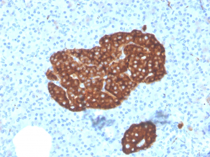
- Scientific DescriptionThis monospecific monoclonal antibody recognizes a protein of 38 kDa that is identified as synaptophysin. It is an N-glycosylated integral membrane protein found in neurons and endocrine cells. Synaptophysin contains four transmembrane domains and may function as a gap junction-like channel. This antibody identifies normal neuroendocrine cells and neuroendocrine neoplasms. Diffuse, finely granular, cytoplasmic staining is observed, which probably correlates with the distribution of the antigen within neurosecretory vesicles. Synaptophysin is an independent, broad-range marker of neural and neuroendocrine differentiation. Primary antibodies are available purified, or with a selection of fluorescent CF® Dyes and other labels. CF® Dyes offer exceptional brightness and photostability. Note: Conjugates of blue fluorescent dyes like CF®405S and CF®405M are not recommended for detecting low abundance targets, because blue dyes have lower fluorescence and can give higher non-specific background than other dye colors.
- SourceAnimal
- ReactivityHuman
- Storage Instruction2°C to 8°C,RT
- UNSPSC41116161
MSDS
Related products
Product group Antibodies
ApplicationsWestern Blot, ELISA, ImmunoHistoChemistry
ReactivityHuman, Mouse, Rat
- SizePrice

Product group Antibodies
Anti-Synaptophysin/SYP Picoband(r) AntibodyA05049-CARRIER-FREE
ApplicationsImmunoFluorescence, Western Blot, ImmunoCytoChemistry, ImmunoHistoChemistry
ReactivityHuman, Mouse, Rat
TargetSYP
- SizePrice
Product group Antibodies

Synaptophysin (SYP) AntibodyABX013199
ApplicationsWestern Blot, ELISA, ImmunoHistoChemistry
- SizePrice
Product group Antibodies
Anti-SYP Antibody144-06344
ApplicationsImmunoFluorescence, Western Blot, ImmunoHistoChemistry
ReactivityHuman, Mouse, Rat
TargetSYP
- SizePrice
Product group Antibodies

ApplicationsImmunoHistoChemistry
ReactivityHuman
TargetSYP
- SizePrice

Product group Antibodies
Synaptophysin Recombinant AntibodyBSM-52379R
ApplicationsFlow Cytometry, ImmunoFluorescence, Western Blot, ImmunoHistoChemistry, ImmunoHistoChemistry Frozen, ImmunoHistoChemistry Paraffin
ReactivityHuman, Mouse, Rat
TargetSYP
- SizePrice
Product group Antibodies
SYP AntibodyCSB-PA004215
ApplicationsWestern Blot, ELISA, ImmunoHistoChemistry
ReactivityHuman, Mouse, Rat
TargetSYP
- SizePrice
Product group Antibodies
ApplicationsImmunoPrecipitation, Western Blot, ImmunoCytoChemistry, ImmunoHistoChemistry
TargetSYP
- SizePrice
![Synaptophysin antibody detects Synaptophysin protein at synaptic vesicles by immunofluorescent analysis. Sample: DIV9 rat E18 primary cortical neurons were fixed in 4% paraformaldehyde at RT for 15 min. Green: Synaptophysin protein stained by Synaptophysin antibody (GTX100865) diluted at 1:500. Red: beta Tubulin 3/ Tuj1, stained by beta Tubulin 3/ Tuj1 antibody [GT11710] (GTX631836) diluted at 1:500. Blue: Fluoroshield with DAPI (GTX30920).](https://www.genetex.com/upload/website/prouct_img/normal/GTX100865/GTX100865_40128_20170503_IFA_R_w_23060100_733.webp)
Product group Antibodies
Synaptophysin antibodyGTX100865
ApplicationsImmunoFluorescence, Western Blot, ImmunoCytoChemistry, ImmunoHistoChemistry, ImmunoHistoChemistry Frozen, ImmunoHistoChemistry Paraffin
ReactivityHuman, Mouse, Rat
TargetSYP
- SizePrice

Product group Antibodies
Anti-SYP AntibodyHPA002858
ApplicationsWestern Blot, ImmunoHistoChemistry
ReactivityHuman
TargetSYP
- SizePrice