

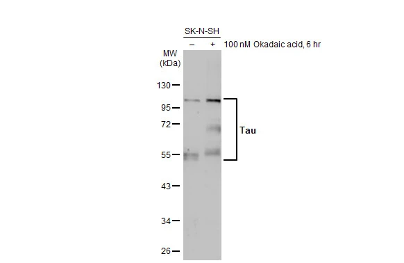
Untreated (–) and treated (+) SK-N-SH whole cell extract (30 μg) were separated by 10% SDS-PAGE, and the membrane was blotted with Tau antibody (GTX130462) diluted at 1:1000. The HRP-conjugated anti-rabbit IgG antibody (GTX213110-01) was used to detect the primary antibody, and the signal was developed with Trident ECL plus-Enhanced.
 was separated by 7.5 % SDS-PAGE, and the membrane was blotted with Tau antibody (GTX130462) at a dilution of 1:10000.")
![Tau antibody detects Tau protein at axon by immunofluorescent analysis. Sample: DIV9 rat E18 primary cortical neurons were fixed in 4% paraformaldehyde at RT for 15 min. Green: Tau protein stained by Tau antibody (GTX130462) diluted at 1:500. Red: MAP2, stained by MAP2 antibody [HM-2] (GTX11267) diluted at 1:1000. Blue: Fluoroshield with DAPI (GTX30920).](https://www.genetex.com/upload/website/prouct_img/normal/GTX130462/GTX130462_41927_20170503_IFA_R_w_23060523_836.webp "Tau antibody detects Tau protein at axon by immunofluorescent analysis. Sample: DIV9 rat E18 primary cortical neurons were fixed in 4% paraformaldehyde at RT for 15 min. Green: Tau protein stained by Tau antibody (GTX130462) diluted at 1:500. Red: MAP2, stained by MAP2 antibody [HM-2] (GTX11267) diluted at 1:1000. Blue: Fluoroshield with DAPI (GTX30920).")
![Tau antibody detects Tau protein expression by immunohistochemical analysis. Sample: Frozen sectioned adult mouse cerebellum. Green: Tau protein stained by Tau antibody (GTX130462) diluted at 1:250. Red: beta Tubulin 3/ TUJ1, a stained by beta Tubulin 3/ TUJ1 antibody [GT11710] (GTX631836) diluted at 1:500. Blue: Fluoroshield with DAPI (GTX30920).](https://www.genetex.com/upload/website/prouct_img/normal/GTX130462/GTX130462_41927_20170622_IHC-Fr_M_w_23060523_486.webp "Tau antibody detects Tau protein expression by immunohistochemical analysis. Sample: Frozen sectioned adult mouse cerebellum. Green: Tau protein stained by Tau antibody (GTX130462) diluted at 1:250. Red: beta Tubulin 3/ TUJ1, a stained by beta Tubulin 3/ TUJ1 antibody [GT11710] (GTX631836) diluted at 1:500. Blue: Fluoroshield with DAPI (GTX30920).")
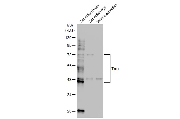
 was separated by 7.5% SDS-PAGE, and the membrane was blotted with Tau antibody (GTX130462) diluted at 1:1000. The HRP-conjugated anti-rabbit IgG antibody (GTX213110-01) was used to detect the primary antibody.")
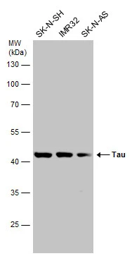
 was separated by 7.5% SDS-PAGE, and the membrane was blotted with Tau antibody (GTX130462) diluted at 1:100000. The HRP-conjugated anti-rabbit IgG antibody (GTX213110-01) was used to detect the primary antibody.")
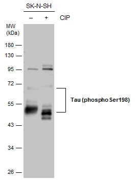
 and transfected (+) SK-N-SH whole cell extracts (30 μg) were separated by 10% SDS-PAGE, and the membrane was blotted with Tau antibody (GTX130462) diluted at 1:500. The HRP-conjugated anti-rabbit IgG antibody (GTX213110-01) was used to detect the primary antibody, and the signal was developed with Trident ECL plus-Enhanced.")
![Tau antibody detects Tau protein at cytoplasm by immunohistochemical analysis. Sample: Paraffin-embedded mouse eye. Green: Tau stained by Tau antibody (GTX130462) diluted at 1:500. Red: beta Tubulin 3/ Tuj1, a cytoskeleton marker, stained by beta Tubulin 3/ Tuj1 antibody [GT11710] (GTX631836) diluted at 1:500. Blue: Fluoroshield with DAPI (GTX30920). Antigen Retrieval: Citrate buffer, pH 6.0, 15 min](https://www.genetex.com/upload/website/prouct_img/normal/GTX130462/GTX130462_41927_20181109_IHC-P-FL_w_23060523_772.webp "Tau antibody detects Tau protein at cytoplasm by immunohistochemical analysis. Sample: Paraffin-embedded mouse eye. Green: Tau stained by Tau antibody (GTX130462) diluted at 1:500. Red: beta Tubulin 3/ Tuj1, a cytoskeleton marker, stained by beta Tubulin 3/ Tuj1 antibody [GT11710] (GTX631836) diluted at 1:500. Blue: Fluoroshield with DAPI (GTX30920). Antigen Retrieval: Citrate buffer, pH 6.0, 15 min")
 was separated by 7.5% SDS-PAGE, and the membranes were blotted with Tau antibody (GTX130462) diluted at 1:4000 and competitor's antibody (#4019) diluted at 1:2000. The HRP-conjugated anti-rabbit IgG antibody (GTX213110-01) was used to detect the primary antibody. *The competitor is not affiliated with GeneTex and does not endorse this product.")
![Tau antibody detects Tau protein at cytoplasm by immunohistochemical analysis. Sample: Paraffin-embedded mouse brain. Green: Tau stained by Tau antibody (GTX130462) diluted at 1:250. Red: alpha Tubulin, a cytoskeleton marker, stained by alpha Tubulin antibody [GT114] (GTX628802) diluted at 1:1000. Blue: Fluoroshield with DAPI (GTX30920). Antigen Retrieval: Citrate buffer, pH 6.0, 15 min](https://www.genetex.com/upload/website/prouct_img/normal/GTX130462/GTX130462_41927_20181005_IHC-P-FL_M_w_23060523_698.webp "Tau antibody detects Tau protein at cytoplasm by immunohistochemical analysis. Sample: Paraffin-embedded mouse brain. Green: Tau stained by Tau antibody (GTX130462) diluted at 1:250. Red: alpha Tubulin, a cytoskeleton marker, stained by alpha Tubulin antibody [GT114] (GTX628802) diluted at 1:1000. Blue: Fluoroshield with DAPI (GTX30920). Antigen Retrieval: Citrate buffer, pH 6.0, 15 min")
Untreated (–) and treated (+) SK-N-SH whole cell extract (30 μg) were separated by 10% SDS-PAGE, and the membrane was blotted with Tau antibody (GTX130462) diluted at 1:1000. The HRP-conjugated anti-rabbit IgG antibody (GTX213110-01) was used to detect the primary antibody, and the signal was developed with Trident ECL plus-Enhanced.
Tau antibody
GTX130462
ApplicationsImmunoFluorescence, Western Blot, ImmunoCytoChemistry, ImmunoHistoChemistry, ImmunoHistoChemistry Frozen, ImmunoHistoChemistry Paraffin
Product group Antibodies
ReactivityHuman, Mouse, Rat
TargetMAPT
Overview
- SupplierGeneTex
- Product NameTau antibody
- Delivery Days Customer9
- Application Supplier NoteWB: 1:500-1:3000. ICC/IF: 1:100-1:1000. IHC-P: 1:100-1:1000. IHC-Fr: 1:100-1:1000. *Optimal dilutions/concentrations should be determined by the researcher.Not tested in other applications.
- ApplicationsImmunoFluorescence, Western Blot, ImmunoCytoChemistry, ImmunoHistoChemistry, ImmunoHistoChemistry Frozen, ImmunoHistoChemistry Paraffin
- CertificationResearch Use Only
- ClonalityPolyclonal
- Concentration0.47 mg/ml
- ConjugateUnconjugated
- Gene ID4137
- Target nameMAPT
- Target descriptionmicrotubule associated protein tau
- Target synonymsDDPAC, FTD1, FTDP-17, MAPTL, MSTD, MTBT1, MTBT2, PPND, PPP1R103, TAU, Tau-PHF6, tau-40, microtubule-associated protein tau, G protein beta1/gamma2 subunit-interacting factor 1, PHF-tau, Tau-derived paired helical filament hexapeptide, neurofibrillary tangle protein, paired helical filament-tau, protein phosphatase 1, regulatory subunit 103
- HostRabbit
- IsotypeIgG
- Protein IDP10636
- Protein NameMicrotubule-associated protein tau
- Scientific DescriptionThis gene encodes the microtubule-associated protein tau (MAPT) whose transcript undergoes complex, regulated alternative splicing, giving rise to several mRNA species. MAPT transcripts are differentially expressed in the nervous system, depending on stage of neuronal maturation and neuron type. MAPT gene mutations have been associated with several neurodegenerative disorders such as Alzheimers disease, Picks disease, frontotemporal dementia, cortico-basal degeneration and progressive supranuclear palsy. [provided by RefSeq]
- ReactivityHuman, Mouse, Rat
- Storage Instruction-20°C or -80°C,2°C to 8°C
- UNSPSC12352203
References
- Li SS, Zhang BY, Yin SG, et al. A new peptide, VD11, promotes structural and functional recovery after spinal cord injury. Neural Regen Res. 2023,18(10):2260-2267. doi: 10.4103/1673-5374.369119Read this paper
- Chen HY, Hsu CL, Lin HY, et al. Clinical and functional characterization of a novel STUB1 frameshift mutation in autosomal dominant spinocerebellar ataxia type 48 (SCA48). J Biomed Sci. 2021,28(1):65. doi: 10.1186/s12929-021-00763-1Read this paper
- Serdar M, Mordelt A, Müser K, et al. Detrimental Impact of Energy Drink Compounds on Developing Oligodendrocytes and Neurons. Cells. 2019,8(11). doi: 10.3390/cells8111381Read this paper
- Shih CH, Chen JK, Kuo LW, et al. Chronic pulmonary exposure to traffic-related fine particulate matter causes brain impairment in adult rats. Part Fibre Toxicol. 2018,15(1):44. doi: 10.1186/s12989-018-0281-1Read this paper
Datasheet
Related products
Product group Antibodies

Anti-Tau [PC1C6]Ab01120-2.0
ApplicationsWestern Blot, ImmunoHistoChemistry
ReactivityBovine, Human, Rat
TargetMAPT
- SizePrice
Product group Antibodies
Anti-Tau (213-222aa) Antibody130-10929
ApplicationsELISA
ReactivityHuman
TargetMAPT
- SizePrice

Product group Antibodies
Anti-Tau/MAPT Antibody Picoband(r)A00097-3-CARRIER-FREE
ApplicationsImmunoFluorescence, Western Blot, ELISA, ImmunoCytoChemistry
ReactivityHuman, Mouse, Rat
TargetMAPT
- SizePrice
Product group Antibodies
References
Tau (phospho Ser198) antibodyGTX130456
ApplicationsWestern Blot
ReactivityHuman, Mouse, Rat
TargetMAPT
- SizePrice
Product group Antibodies
References
Tau antibodyGTX116044
ApplicationsImmunoFluorescence, Western Blot, ImmunoCytoChemistry
ReactivityHuman
TargetMAPT
- SizePrice
Product group Antibodies
References
Tau (phospho Thr231) antibodyGTX01563
ApplicationsWestern Blot
ReactivityHuman, Mouse, Rat
TargetMAPT
- SizePrice
![IHC-P analysis of rat brain tissue section using GTX03204 Tau (phospho Ser396) antibody [GT1292]. Dilution : 1:100](https://www.genetex.com/upload/website/prouct_img/normal/GTX03204/GTX03204_20210615_IHC-P_89_w_23053123_804.webp)
Product group Antibodies
ApplicationsWestern Blot, ImmunoHistoChemistry, ImmunoHistoChemistry Paraffin
ReactivityHuman, Mouse, Rat
TargetMAPT
- SizePrice
Product group Antibodies
ApplicationsWestern Blot
ReactivityHuman
TargetMAPT
- SizePrice