

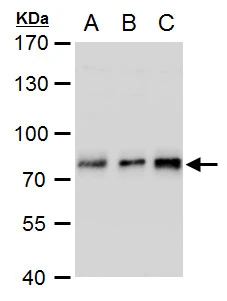
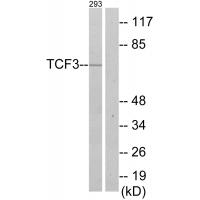
TCF3 / E2A antibody detects TCF3 protein by western blot analysis. A. 30 μg Jurkat whole cell extract B. 30 μg Raji whole cell extract C. 30 μg NCI-H929 whole cell extract 7.5 % SDS-PAGE TCF3 / E2A antibody (GTX129237) dilution: 1:2500
 dilution: 1:3000")
 diluted at 1:500. Blue: Hoechst 33342 staining.")
![TCF3 / E2A antibody immunoprecipitates TCF3 protein in IP experiments. IP samples: Jurkat whole cell extract A. 50 μg Jurkat whole cell extract B. Control with 4 μg of preimmune Rabbit IgG C. Immunoprecipitation of TCF3 protein by 4 μg TCF3 / E2A antibody (GTX129237) 7.5 % SDS-PAGE The immunoprecipitated TCF3 protein was detected by TCF3 / E2A antibody (GTX129237) diluted at 1:500. [EasyBlot anti-rabbit IgG (GTX221666-01) was used as a secondary reagent]](https://www.genetex.com/upload/website/prouct_img/normal/GTX129237/GTX129237_41626_IP_w_23060523_602.webp "TCF3 / E2A antibody immunoprecipitates TCF3 protein in IP experiments. IP samples: Jurkat whole cell extract A. 50 μg Jurkat whole cell extract B. Control with 4 μg of preimmune Rabbit IgG C. Immunoprecipitation of TCF3 protein by 4 μg TCF3 / E2A antibody (GTX129237) 7.5 % SDS-PAGE The immunoprecipitated TCF3 protein was detected by TCF3 / E2A antibody (GTX129237) diluted at 1:500. [EasyBlot anti-rabbit IgG (GTX221666-01) was used as a secondary reagent]")
TCF3 / E2A antibody detects TCF3 protein by western blot analysis. A. 30 μg Jurkat whole cell extract B. 30 μg Raji whole cell extract C. 30 μg NCI-H929 whole cell extract 7.5 % SDS-PAGE TCF3 / E2A antibody (GTX129237) dilution: 1:2500
TCF3 / E2A antibody
GTX129237
ApplicationsImmunoFluorescence, ImmunoPrecipitation, Western Blot, ImmunoCytoChemistry
Product group Antibodies
ReactivityHuman
TargetTCF3
Overview
- SupplierGeneTex
- Product NameTCF3 / E2A antibody
- Delivery Days Customer9
- Application Supplier NoteWB: 1:1000-1:3000. ICC/IF: 1:100-1:1000. IP: 1:100-1:500. *Optimal dilutions/concentrations should be determined by the researcher.Not tested in other applications.
- ApplicationsImmunoFluorescence, ImmunoPrecipitation, Western Blot, ImmunoCytoChemistry
- CertificationResearch Use Only
- ClonalityPolyclonal
- Concentration1 mg/ml
- ConjugateUnconjugated
- Gene ID6929
- Target nameTCF3
- Target descriptiontranscription factor 3
- Target synonymsAGM8, AGM8A, AGM8B, E2A, E47, ITF1, TCF-3, VDIR, bHLHb21, p75, transcription factor E2-alpha, E2A-HLF fusion transcript protein, NOL1-TCF3 fusion, VDR interacting repressor, class B basic helix-loop-helix protein 21, helix-loop-helix protein HE47, immunoglobulin transcription factor 1, kappa-E2-binding factor, negative vitamin D response element-binding protein, transcription factor 3 (E2A immunoglobulin enhancer binding factors E12/E47), transcription factor ITF-1, vitamin D receptor-interacting repressor
- HostRabbit
- IsotypeIgG
- Protein IDP15923
- Protein NameTranscription factor E2-alpha
- Scientific DescriptionThe TCF3 gene, also called E2A, encodes 2 basic helix-loop-helix (bHLH) transcription factors, E12 and E47, through alternative splicing. E12 and E47 are involved in regulation of immunoglobulin gene expression (Bain et al., 1994 [PubMed 8001125]).[supplied by OMIM]
- ReactivityHuman
- Storage Instruction-20°C or -80°C,2°C to 8°C
- UNSPSC12352203
Datasheet
Related products
Product group Antibodies
Anti-TCF3 Antibody101-11755
ApplicationsWestern Blot, ELISA
TargetTCF3
- SizePrice

Product group Antibodies
Anti-Phospho-E2A (T355) TCF3 AntibodyA00095T355
ApplicationsImmunoFluorescence, ELISA, ImmunoHistoChemistry
ReactivityHuman, Mouse, Rat
TargetTCF3
- SizePrice
Product group Antibodies
TCF3 / E2A antibodyGTX109885
ApplicationsImmunoFluorescence, ImmunoPrecipitation, Western Blot, ImmunoCytoChemistry, ImmunoHistoChemistry, ImmunoHistoChemistry Paraffin
ReactivityHuman, Mouse, Rat
TargetTCF3
- SizePrice
Product group Antibodies
TCF3 / E2A antibodyGTX110458
ApplicationsImmunoFluorescence, Western Blot, ImmunoCytoChemistry
ReactivityHuman
TargetTCF3
- SizePrice
![TCF3 / E2A antibody [HL1954] detects TCF3 / E2A protein by immunofluorescent analysis. Sample: SH-SY-5Y cells were fixed in 4% paraformaldehyde at RT for 15 min. Green: TCF3 / E2A stained by TCF3 / E2A antibody [HL1954] (GTX637791) diluted at 1:500. Red: alpha Tubulin, a cytoskeleton marker, stained by alpha Tubulin antibody [GT114] (GTX628802) diluted at 1:1000. Blue: Fluoroshield with DAPI (GTX30920). Scale bar= 10μm.](https://www.genetex.com/upload/website/prouct_img/normal/GTX637791/GTX637791_44900_20221230_ICC_IF_22122901_734.webp)
Product group Antibodies
TCF3 / E2A antibody [HL1954]GTX637791
ApplicationsImmunoFluorescence, Western Blot, ImmunoCytoChemistry
ReactivityHuman
TargetTCF3
- SizePrice
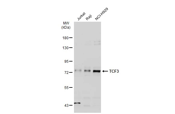
![Various whole cell extracts (30 μg) were separated by 7.5% SDS-PAGE, and the membrane was blotted with TCF3 / E2A antibody [HL1955] (GTX637792) diluted at 1:1000. The HRP-conjugated anti-rabbit IgG antibody (GTX213110-01) was used to detect the primary antibody, and the signal was developed with Trident ECL plus-Enhanced.](https://www.genetex.com/upload/website/prouct_img/normal/GTX637792/GTX637792_44900_20221230_WB_23010400_261.webp)
Product group Antibodies
TCF3 / E2A antibody [HL1955]GTX637792
ApplicationsImmunoFluorescence, Western Blot, ImmunoCytoChemistry
ReactivityHuman
TargetTCF3
- SizePrice
Product group Antibodies
TCF3 Recombinant AntibodyBSM-60695R
ApplicationsImmunoFluorescence, Western Blot, ImmunoHistoChemistry, ImmunoHistoChemistry Frozen, ImmunoHistoChemistry Paraffin
ReactivityHuman
TargetTCF3
- SizePrice
Product group Antibodies
TCF3 AntibodyCSB-PA004248
ApplicationsWestern Blot, ELISA, ImmunoHistoChemistry
ReactivityHuman, Mouse, Rat
TargetTCF3
- SizePrice
Product group Antibodies
Anti-TCF3 AntibodyA38611
ApplicationsWestern Blot, ImmunoHistoChemistry
ReactivityHuman, Mouse, Rat
- SizePrice
Product group Antibodies
TCF3 / E2A antibody, InternalGTX89171
ApplicationsWestern Blot, ImmunoHistoChemistry, ImmunoHistoChemistry Paraffin
ReactivityHuman
TargetTCF3
- SizePrice