


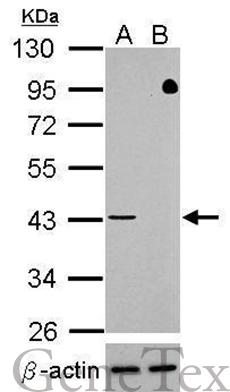
Wild-type (WT) and TARDBP knockout (KO) HAP1 cell extracts (50 Aμg) were separated by gradient gel% SDS-PAGE, and the membrane was blotted with TDP43 antibody [GT733] (GTX630197) diluted at 1:500. The HRP-conjugated anti-mouse IgG antibody (GTX213111-01) was used to detect the primary antibody. Data provided byA?YCharOSA?Inc., an open science company with the mission of characterizing commercially available antibody reagents for all human proteins using knockout technology.
![TDP43 antibody [GT733] detects TDP43 protein at nucleus in mouse brain by immunohistochemical analysis. Sample: Paraffin-embedded mouse brain. TDP43 antibody [GT733] (GTX630197) diluted at 1:200.
Antigen Retrieval: Citrate buffer, pH 6.0, 15 min](https://www.genetex.com/upload/website/prouct_img/normal/GTX630197/GTX630197_41505_20171020_IHC-P_M_w_23061202_225.webp "TDP43 antibody [GT733] detects TDP43 protein at nucleus in mouse brain by immunohistochemical analysis. Sample: Paraffin-embedded mouse brain. TDP43 antibody [GT733] (GTX630197) diluted at 1:200.
Antigen Retrieval: Citrate buffer, pH 6.0, 15 min")
![Various whole cell extracts (30 μg) were separated by 10% SDS-PAGE, and the membrane was blotted with TARDBP antibody [GT733] (GTX630197) diluted at 1:1000. The signal was developed with Trident ECL plus-Enhanced.](https://www.genetex.com/upload/website/prouct_img/normal/GTX630197/GTX630197_41505_20160505_WB_w_23061202_745.webp "Various whole cell extracts (30 μg) were separated by 10% SDS-PAGE, and the membrane was blotted with TARDBP antibody [GT733] (GTX630197) diluted at 1:1000. The signal was developed with Trident ECL plus-Enhanced.")
![TARDBP antibody [GT733] validation by siRNA knock-down. Upperpanel: TARDBP antibody [GT733] GTX630197 Lower panel: GAPDH antibody (GTX100118) A. 30 μg 293T whole cell lysate/extract B. 30 μg whole cell lysate/extract of TARDBP shRNA-transfected 293T cells 10 % SDS-PAGE TARDBP antibody [GT733] (GTX630197) dilution: 1:1000 GAPDH antibody (GTX100118) dilution: 1:10000](https://www.genetex.com/upload/website/prouct_img/normal/GTX630197/GTX630197_41505_WB_shRNA_watermark_w_23061202_231.webp "TARDBP antibody [GT733] validation by siRNA knock-down. Upperpanel: TARDBP antibody [GT733] GTX630197 Lower panel: GAPDH antibody (GTX100118) A. 30 μg 293T whole cell lysate/extract B. 30 μg whole cell lysate/extract of TARDBP shRNA-transfected 293T cells 10 % SDS-PAGE TARDBP antibody [GT733] (GTX630197) dilution: 1:1000 GAPDH antibody (GTX100118) dilution: 1:10000")
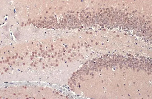
![TDP43 antibody [GT733] detects TDP43 protein at nucleus in rat brain by immunohistochemical analysis. Sample: Paraffin-embedded rat brain. TDP43 antibody [GT733] (GTX630197) diluted at 1:200.
Antigen Retrieval: Citrate buffer, pH 6.0, 15 min](https://www.genetex.com/upload/website/prouct_img/normal/GTX630197/GTX630197_41505_20171020_IHC-P_R_w_23061202_203.webp "TDP43 antibody [GT733] detects TDP43 protein at nucleus in rat brain by immunohistochemical analysis. Sample: Paraffin-embedded rat brain. TDP43 antibody [GT733] (GTX630197) diluted at 1:200.
Antigen Retrieval: Citrate buffer, pH 6.0, 15 min")
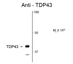
![TARDBP antibody [GT733] detects TARDBP protein by western blot analysis. A. 30 μg 293T whole cell lysate/extract B. 30 μg A431 whole cell lysate/extract C. 30 μg HeLa whole cell lysate/extract D. 30 μg HepG2 whole cell lysate/extract 10 % SDS-PAGE TARDBP antibody [GT733] (GTX630197) dilution: 1:1000](https://www.genetex.com/upload/website/prouct_img/normal/GTX630197/GTX630197_41505_WB_w_23061202_538.webp "TARDBP antibody [GT733] detects TARDBP protein by western blot analysis. A. 30 μg 293T whole cell lysate/extract B. 30 μg A431 whole cell lysate/extract C. 30 μg HeLa whole cell lysate/extract D. 30 μg HepG2 whole cell lysate/extract 10 % SDS-PAGE TARDBP antibody [GT733] (GTX630197) dilution: 1:1000")
![TDP43 antibody [GT733] detects TDP43 protein at nucleus by immunofluorescent analysis. Sample: HeLa cells were fixed in 4% paraformaldehyde at RT for 15 min. Green: TDP43 stained by TDP43 antibody [GT733] (GTX630197) diluted at 1:500. Red: beta Tubulin, a cytoskeleton marker, stained by beta Tubulin antibody (GTX101279) diluted at 1:1000.](https://www.genetex.com/upload/website/prouct_img/normal/GTX630197/GTX630197_45327_20240315_ICC_IF_24041019_101.webp "TDP43 antibody [GT733] detects TDP43 protein at nucleus by immunofluorescent analysis. Sample: HeLa cells were fixed in 4% paraformaldehyde at RT for 15 min. Green: TDP43 stained by TDP43 antibody [GT733] (GTX630197) diluted at 1:500. Red: beta Tubulin, a cytoskeleton marker, stained by beta Tubulin antibody (GTX101279) diluted at 1:1000.")
Wild-type (WT) and TARDBP knockout (KO) HAP1 cell extracts (50 Aμg) were separated by gradient gel% SDS-PAGE, and the membrane was blotted with TDP43 antibody [GT733] (GTX630197) diluted at 1:500. The HRP-conjugated anti-mouse IgG antibody (GTX213111-01) was used to detect the primary antibody. Data provided byA?YCharOSA?Inc., an open science company with the mission of characterizing commercially available antibody reagents for all human proteins using knockout technology.
TDP43 antibody [GT733]
GTX630197
ApplicationsImmunoFluorescence, ImmunoPrecipitation, Western Blot, ImmunoCytoChemistry, ImmunoHistoChemistry, ImmunoHistoChemistry Paraffin
Product group Antibodies
ReactivityHuman, Mouse, Rat
TargetTARDBP
Overview
- SupplierGeneTex
- Product NameTDP43 antibody [GT733]
- Delivery Days Customer9
- Application Supplier NoteWB: 1:500-1:3000. ICC/IF: 1:100-1:1000. IHC-P: 1:100-1:1000. *Optimal dilutions/concentrations should be determined by the researcher.Not tested in other applications.
- ApplicationsImmunoFluorescence, ImmunoPrecipitation, Western Blot, ImmunoCytoChemistry, ImmunoHistoChemistry, ImmunoHistoChemistry Paraffin
- CertificationResearch Use Only
- ClonalityMonoclonal
- Clone IDGT733
- Concentration1 mg/ml
- ConjugateUnconjugated
- Gene ID23435
- Target nameTARDBP
- Target descriptionTAR DNA binding protein
- Target synonymsALS10, TDP-43, TAR DNA-binding protein 43, TAR DNA-binding protein-43
- HostMouse
- IsotypeIgG1
- Protein IDQ13148
- Protein NameTAR DNA-binding protein 43
- Scientific DescriptionHIV-1, the causative agent of acquired immunodeficiency syndrome (AIDS), contains an RNA genome that produces a chromosomally integrated DNA during the replicative cycle. Activation of HIV-1 gene expression by the transactivator Tat is dependent on an RNA regulatory element (TAR) located downstream of the transcription initiation site. The protein encoded by this gene is a transcriptional repressor that binds to chromosomally integrated TAR DNA and represses HIV-1 transcription. In addition, this protein regulates alternate splicing of the CFTR gene. A similar pseudogene is present on chromosome 20. [provided by RefSeq, Jul 2008]
- ReactivityHuman, Mouse, Rat
- Storage Instruction-20°C or -80°C,2°C to 8°C
- UNSPSC12352203
References
- Worrall D, Ayoubi R, Fotouhi M, et al. The identification of high-performing antibodies for TDP-43 for use in Western Blot, immunoprecipitation and immunofluorescence. F1000Res. 2023,12:277. doi: 10.12688/f1000research.131852.2Read this paper
Datasheet
Related products
Product group Antibodies
Anti-TARDBP Antibody144-01183
ApplicationsImmunoFluorescence, ImmunoPrecipitation, Western Blot, ImmunoHistoChemistry, Other Application
ReactivityHuman, Mouse, Rat
TargetTARDBP
- SizePrice
Product group Antibodies
TDP43 antibodyGTX100579
ApplicationsImmunoFluorescence, Western Blot, ImmunoCytoChemistry, ImmunoHistoChemistry, ImmunoHistoChemistry Paraffin
ReactivityHuman, Mouse, Rat
TargetTARDBP
- SizePrice
Product group Antibodies
References
TDP43 antibodyGTX114210
ApplicationsImmunoFluorescence, ImmunoPrecipitation, Western Blot, ChIP Chromatin ImmunoPrecipitation, ImmunoCytoChemistry, ImmunoHistoChemistry, ImmunoHistoChemistry Paraffin, Other Application
ReactivityHuman, Mouse, Rat
TargetTARDBP
- SizePrice
Product group Antibodies
TDP43 antibody [k1B9]GTX57560
ApplicationsFlow Cytometry, Western Blot, ImmunoHistoChemistry, ImmunoHistoChemistry Paraffin
ReactivityHuman
TargetTARDBP
- SizePrice
![Wild-type (WT) and TARDBP knockout (KO) HAP1 cell extracts (50 Aμg) were separated by gradient gel% SDS-PAGE, and the membrane was blotted with TDP43 antibody [GT225] (GTX630196) diluted at 1:500. The HRP-conjugated anti-mouse IgG antibody (GTX213111-01) was used to detect the primary antibody. Data provided byA?YCharOSA?Inc., an open science company with the mission of characterizing commercially available antibody reagents for all human proteins using knockout technology.](https://www.genetex.com/upload/website/prouct_img/normal/GTX630196/GTX630196_41505_20220805_WB_KO_22080923_561.webp)
Product group Antibodies
References
TDP43 antibody [GT225]GTX630196
ApplicationsWestern Blot, ImmunoHistoChemistry, ImmunoHistoChemistry Paraffin
ReactivityHuman, Mouse, Rat
TargetTARDBP
- SizePrice
![TDP43 antibody [GT6310] detects TDP43 Protein expression by immunohistochemical analysis. Sample: Frozen-sectioned adult mouse cerebellum. Green: TDP43 stained by TDP43 antibody [GT6310] (GTX633973) diluted at 1:250. Red: Calbindin, stained by Calbindin antibody (GTX130856) diluted at 1:500. Blue: Fluoroshield with DAPI (GTX30920).
Antigen Retrieval: Citrate buffer, pH 6.0, 5 min](https://www.genetex.com/upload/website/prouct_img/normal/GTX633973/GTX633973_42730_20170829_IHC-Fr_M_w_23061202_828.webp)
Product group Antibodies
TDP43 antibody [GT6310]GTX633973
ApplicationsWestern Blot, ImmunoHistoChemistry, ImmunoHistoChemistry Frozen, ImmunoHistoChemistry Paraffin
ReactivityHuman, Mouse, Rat
TargetTARDBP
- SizePrice
Product group Antibodies
References
TDP43 antibodyGTX82580
ApplicationsWestern Blot, ImmunoHistoChemistry
ReactivityHuman, Rat
TargetTARDBP
- SizePrice

Product group Antibodies
Anti-TDP-43/TARDBP Antibody Picoband(r)A01001-3-CARRIER-FREE
ApplicationsFlow Cytometry, ImmunoFluorescence, Western Blot, ELISA, ImmunoCytoChemistry, ImmunoHistoChemistry
ReactivityHuman, Mouse, Rat
TargetTARDBP
- SizePrice
Product group Antibodies
TARDBP Polyclonal AntibodyCAC14898
ApplicationsImmunoFluorescence, Western Blot, ELISA, ImmunoHistoChemistry
TargetTARDBP
- SizePrice