

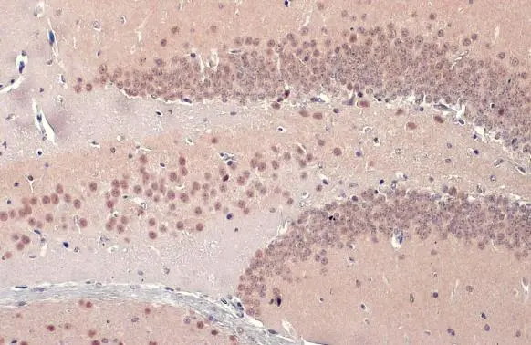
TDP43 antibody detects TDP43 protein at nucleus by immunohistochemical analysis. Sample: Paraffin-embedded mouse brain. TDP43 stained by TDP43 antibody (GTX114210) diluted at 1:500. Antigen Retrieval: Citrate buffer, pH 6.0, 15 min
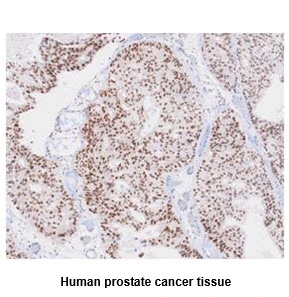
 antibody at 1:100 dilution.
Antigen Retrieval: Trilogy? (EDTA based, pH 8.0) buffer, 15min")
 diluted at 1:2000. Red: phalloidin, a cytoskeleton marker, diluted at 1:200.")
 12% SDS-PAGE The immunoprecipitated TARDBP protein was detected by TARDBP antibody (GTX114210) diluted at 1:1000. EasyBlot anti-rabbit IgG (GTX221666-01) was used as a secondary reagent.")
 diluted at 1:500. Antigen Retrieval: Citrate buffer, pH 6.0, 15 min")
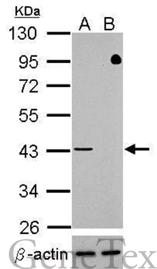
 were separated by 10% SDS-PAGE, and the membrane was blotted with TDP43 antibody (GTX114210) diluted at 1:2000. The HRP-conjugated anti-rabbit IgG antibody (GTX213110-01) was used to detect the primary antibody.")
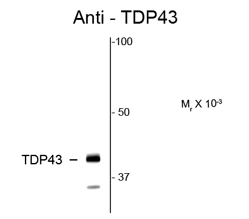
 dilution: 1:3000 The HRP-conjugated anti-rabbit IgG antibody (GTX213110-01) was used to detect the primary antibody.")
![TDP43 antibody detects TDP43 protein by immunofluorescent analysis. Sample: DIV10 rat E18 primary cortical neuron cells were fixed in 4% paraformaldehyde at RT for 15 min. Green: TDP43 stained by TDP43 antibody (GTX114210) diluted at 1:500. Red: Tau, stained by Tau antibody [GT287] (GTX634809) diluted at 1:500. Blue: Fluoroshield with DAPI (GTX30920).](https://www.genetex.com/upload/website/prouct_img/normal/GTX114210/GTX114210_43670_20191125_ICC_IF_R_w_23060501_537.webp "TDP43 antibody detects TDP43 protein by immunofluorescent analysis. Sample: DIV10 rat E18 primary cortical neuron cells were fixed in 4% paraformaldehyde at RT for 15 min. Green: TDP43 stained by TDP43 antibody (GTX114210) diluted at 1:500. Red: Tau, stained by Tau antibody [GT287] (GTX634809) diluted at 1:500. Blue: Fluoroshield with DAPI (GTX30920).")
 were separated by 10% SDS-PAGE, and the membrane was blotted with TDP43 antibody (GTX114210) diluted at 1:1000. The HRP-conjugated anti-rabbit IgG antibody (GTX213110-01) was used to detect the primary antibody.")
TDP43 antibody detects TDP43 protein at nucleus by immunohistochemical analysis. Sample: Paraffin-embedded mouse brain. TDP43 stained by TDP43 antibody (GTX114210) diluted at 1:500. Antigen Retrieval: Citrate buffer, pH 6.0, 15 min
TDP43 antibody
GTX114210
ApplicationsImmunoFluorescence, ImmunoPrecipitation, Western Blot, ChIP Chromatin ImmunoPrecipitation, ImmunoCytoChemistry, ImmunoHistoChemistry, ImmunoHistoChemistry Paraffin, Other Application
Product group Antibodies
ReactivityHuman, Mouse, Rat
TargetTARDBP
Overview
- SupplierGeneTex
- Product NameTDP43 antibody
- Delivery Days Customer9
- Application Supplier NoteWB: 1:500-1:3000. ICC/IF: 1:100-1:1000. IHC-P: 1:100-1:1000. IP: 1:100-1:1000. *Optimal dilutions/concentrations should be determined by the researcher.Not tested in other applications.
- ApplicationsImmunoFluorescence, ImmunoPrecipitation, Western Blot, ChIP Chromatin ImmunoPrecipitation, ImmunoCytoChemistry, ImmunoHistoChemistry, ImmunoHistoChemistry Paraffin, Other Application
- CertificationResearch Use Only
- ClonalityPolyclonal
- Concentration0.63 mg/ml
- ConjugateUnconjugated
- Gene ID23435
- Target nameTARDBP
- Target descriptionTAR DNA binding protein
- Target synonymsALS10, TDP-43, TAR DNA-binding protein 43, TAR DNA-binding protein-43
- HostRabbit
- IsotypeIgG
- Protein IDQ13148
- Protein NameTAR DNA-binding protein 43
- Scientific DescriptionHIV-1, the causative agent of acquired immunodeficiency syndrome (AIDS), contains an RNA genome that produces a chromosomally integrated DNA during the replicative cycle. Activation of HIV-1 gene expression by the transactivator Tat is dependent on an RNA regulatory element (TAR) located downstream of the transcription initiation site. The protein encoded by this gene is a transcriptional repressor that binds to chromosomally integrated TAR DNA and represses HIV-1 transcription. In addition, this protein regulates alternate splicing of the CFTR gene. A similar pseudogene is present on chromosome 20. [provided by RefSeq]
- ReactivityHuman, Mouse, Rat
- Storage Instruction-20°C or -80°C,2°C to 8°C
- UNSPSC12352203
References
- Harrell TL, Davido DJ, Bertke AS. Herpes Simplex Virus 1 (HSV-1) Infected Cell Protein 0 (ICP0) Targets of Ubiquitination during Productive Infection of Primary Adult Sensory Neurons. Int J Mol Sci. 2023,24(3). doi: 10.3390/ijms24032931Read this paper
- Chu JF, Majumder P, Chatterjee B, et al. TDP-43 Regulates Coupled Dendritic mRNA Transport-Translation Processes in Co-operation with FMRP and Staufen1. Cell Rep. 2019,29(10):3118-3133.e6. doi: 10.1016/j.celrep.2019.10.061Read this paper
- Wu LS, Cheng WC, Chen CY, et al. Transcriptomopathies of pre- and post-symptomatic frontotemporal dementia-like mice with TDP-43 depletion in forebrain neurons. Acta Neuropathol Commun. 2019,7(1):50. doi: 10.1186/s40478-019-0674-xRead this paper
- Majumder P, Chu JF, Chatterjee B, et al. Co-regulation of mRNA translation by TDP-43 and Fragile X Syndrome protein FMRP. Acta Neuropathol. 2016,132(5):721-738.Read this paper
- Sundararaman B, Zhan L, Blue SM, et al. Resources for the Comprehensive Discovery of Functional RNA Elements. Mol Cell. 2016,61(6):903-13. doi: 10.1016/j.molcel.2016.02.012Read this paper
- Huang CC, Bose JK, Majumder P, et al. Metabolism and mis-metabolism of the neuropathological signature protein TDP-43. J Cell Sci. 2014,127(Pt 14):3024-38. doi: 10.1242/jcs.136150Read this paper
Datasheet
Related products
Product group Antibodies
Anti-TARDBP Antibody144-01183
ApplicationsImmunoFluorescence, ImmunoPrecipitation, Western Blot, ImmunoHistoChemistry, Other Application
ReactivityHuman, Mouse, Rat
TargetTARDBP
- SizePrice
Product group Antibodies
TDP43 antibodyGTX100579
ApplicationsImmunoFluorescence, Western Blot, ImmunoCytoChemistry, ImmunoHistoChemistry, ImmunoHistoChemistry Paraffin
ReactivityHuman, Mouse, Rat
TargetTARDBP
- SizePrice
Product group Antibodies
TDP43 antibody [k1B9]GTX57560
ApplicationsFlow Cytometry, Western Blot, ImmunoHistoChemistry, ImmunoHistoChemistry Paraffin
ReactivityHuman
TargetTARDBP
- SizePrice
![Wild-type (WT) and TARDBP knockout (KO) HAP1 cell extracts (50 Aμg) were separated by gradient gel% SDS-PAGE, and the membrane was blotted with TDP43 antibody [GT225] (GTX630196) diluted at 1:500. The HRP-conjugated anti-mouse IgG antibody (GTX213111-01) was used to detect the primary antibody. Data provided byA?YCharOSA?Inc., an open science company with the mission of characterizing commercially available antibody reagents for all human proteins using knockout technology.](https://www.genetex.com/upload/website/prouct_img/normal/GTX630196/GTX630196_41505_20220805_WB_KO_22080923_561.webp)
Product group Antibodies
References
TDP43 antibody [GT225]GTX630196
ApplicationsWestern Blot, ImmunoHistoChemistry, ImmunoHistoChemistry Paraffin
ReactivityHuman, Mouse, Rat
TargetTARDBP
- SizePrice
![Wild-type (WT) and TARDBP knockout (KO) HAP1 cell extracts (50 Aμg) were separated by gradient gel% SDS-PAGE, and the membrane was blotted with TDP43 antibody [GT733] (GTX630197) diluted at 1:500. The HRP-conjugated anti-mouse IgG antibody (GTX213111-01) was used to detect the primary antibody. Data provided byA?YCharOSA?Inc., an open science company with the mission of characterizing commercially available antibody reagents for all human proteins using knockout technology.](https://www.genetex.com/upload/website/prouct_img/normal/GTX630197/GTX630197_41505_20220805_WB_KO_22080923_274.webp)
Product group Antibodies
References
TDP43 antibody [GT733]GTX630197
ApplicationsImmunoFluorescence, ImmunoPrecipitation, Western Blot, ImmunoCytoChemistry, ImmunoHistoChemistry, ImmunoHistoChemistry Paraffin
ReactivityHuman, Mouse, Rat
TargetTARDBP
- SizePrice
![TDP43 antibody [GT6310] detects TDP43 Protein expression by immunohistochemical analysis. Sample: Frozen-sectioned adult mouse cerebellum. Green: TDP43 stained by TDP43 antibody [GT6310] (GTX633973) diluted at 1:250. Red: Calbindin, stained by Calbindin antibody (GTX130856) diluted at 1:500. Blue: Fluoroshield with DAPI (GTX30920).
Antigen Retrieval: Citrate buffer, pH 6.0, 5 min](https://www.genetex.com/upload/website/prouct_img/normal/GTX633973/GTX633973_42730_20170829_IHC-Fr_M_w_23061202_828.webp)
Product group Antibodies
TDP43 antibody [GT6310]GTX633973
ApplicationsWestern Blot, ImmunoHistoChemistry, ImmunoHistoChemistry Frozen, ImmunoHistoChemistry Paraffin
ReactivityHuman, Mouse, Rat
TargetTARDBP
- SizePrice
Product group Antibodies
References
TDP43 antibodyGTX82580
ApplicationsWestern Blot, ImmunoHistoChemistry
ReactivityHuman, Rat
TargetTARDBP
- SizePrice

Product group Antibodies
Anti-TDP-43/TARDBP Antibody Picoband(r)A01001-3-CARRIER-FREE
ApplicationsFlow Cytometry, ImmunoFluorescence, Western Blot, ELISA, ImmunoCytoChemistry, ImmunoHistoChemistry
ReactivityHuman, Mouse, Rat
TargetTARDBP
- SizePrice
Product group Antibodies
TARDBP Polyclonal AntibodyCAC14898
ApplicationsImmunoFluorescence, Western Blot, ELISA, ImmunoHistoChemistry
TargetTARDBP
- SizePrice