

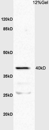
L1 mouse intestine lysates probed with Anti TNFR1/TNF Receptor I Polyclonal Antibody, Unconjugated (bs-2941R) at 1:200 overnight at 4˚C. Followed by conjugation to secondary antibody (bs-0295G-HRP) at 1:3000 for 90 min at 37˚C. Predicted band 40kD/26kD. Observed band size:40kD/26kD.\n
 were incubated in 5% BSA blocking buffer for 30 min at room temperature. Cells were then stained with TNF Antibody(bs-2941R) at 1:50 dilution in blocking buffer and incubated for 30 min at room temperature, washed twice with 2% BSA in PBS. Acquisitions of 20,000 events were performed. Cells stained with primary antibody (green) and isotype control (orange).")
 at 1:1000 dilution and 4˚C overnight incubation. Followed by conjugated secondary antibody incubation at 1:20000 for 60 min at 37˚C.")
at 1:100 dilution in blocking buffer and incubated for 30 min at room temperature, washed twice with 2%BSA in PBS, followed by secondary antibody incubation for 40 min at room temperature. Acquisitions of 20,000 events were performed. Cells stained with primary antibody (green), and isotype control (orange).")
L1 mouse intestine lysates probed with Anti TNFR1/TNF Receptor I Polyclonal Antibody, Unconjugated (bs-2941R) at 1:200 overnight at 4˚C. Followed by conjugation to secondary antibody (bs-0295G-HRP) at 1:3000 for 90 min at 37˚C. Predicted band 40kD/26kD. Observed band size:40kD/26kD.\n
TNFR1 Polyclonal Antibody
BS-2941R
ApplicationsFlow Cytometry, ImmunoFluorescence, Western Blot, ELISA, ImmunoCytoChemistry, ImmunoHistoChemistry, ImmunoHistoChemistry Paraffin
Product group Antibodies
ReactivityBovine, Canine, Equine, Human, Mouse, Porcine, Rabbit, Rat
TargetTNFRSF1A
Overview
- SupplierBioss
- Product NameTNFR1 Polyclonal Antibody
- Delivery Days Customer16
- ApplicationsFlow Cytometry, ImmunoFluorescence, Western Blot, ELISA, ImmunoCytoChemistry, ImmunoHistoChemistry, ImmunoHistoChemistry Paraffin
- Applications SupplierWB(1:300-5000), ELISA(1:500-1000), FCM(1:20-100), IHC-P(1:200-400), IF(ICC)(1:50-200)
- CertificationResearch Use Only
- ClonalityPolyclonal
- Concentration1 ug/ul
- ConjugateUnconjugated
- Gene ID7132
- Target nameTNFRSF1A
- Target descriptionTNF receptor superfamily member 1A
- Target synonymsCD120a, FPF, TBP1, TNF-R, TNF-R-I, TNF-R55, TNFAR, TNFR1, TNFR55, TNFR60, p55, p55-R, p60, tumor necrosis factor receptor superfamily member 1A, TNF-R1, TNF-RI, TNFR-I, tumor necrosis factor binding protein 1, tumor necrosis factor receptor type 1, tumor necrosis factor-alpha receptor
- HostRabbit
- IsotypeIgG
- Protein IDP19438
- Protein NameTumor necrosis factor receptor superfamily member 1A
- ReactivityBovine, Canine, Equine, Human, Mouse, Porcine, Rabbit, Rat
- Storage Instruction-20°C
- UNSPSC41116161
References
- Tumor necrosis factor alpha inhibitors have no effect on a human T-lymphotropic virus type-I (HTLV-I)-infected cell line from patients with HTLV-I-associated myelopathy. Fukui S et al., 2017 Feb 3, BMC ImmunolRead this paper
- Macrolide antibiotics differentially influence human HepG2 cytotoxicity and modulate intrinsic/extrinsic apoptotic pathways in rat hepatocellular carcinoma model. Abdel-Hamid NI et al., 2017 Apr, Naunyn Schmiedebergs Arch PharmacolRead this paper
- Inhibitory effect of Zanthoxylum bungeanum seed oil on ovalbumin-induced lung inflammation in a murine model of asthma. Wang JQ et al., 2016 May, Mol Med RepRead this paper
Datasheet
Related products
Product group Antibodies
Anti-TNFRSF1A AntibodyA49067
ApplicationsWestern Blot, ELISA
ReactivityHuman
- SizePrice
Product group Antibodies

TNFR-S274 AntibodyABX026117
ApplicationsFlow Cytometry, Western Blot, ELISA
- SizePrice
Product group Antibodies

ApplicationsELISA
ReactivityHuman
TargetTNFRSF1A
- SizePrice

Product group Antibodies
Anti-TNF Receptor I/TNFRSF1A Antibody Picoband(r)A00294-3-CARRIER-FREE
ApplicationsFlow Cytometry, Western Blot, ELISA, ImmunoHistoChemistry
ReactivityHuman, Mouse, Rat
TargetTNFRSF1A
- SizePrice
Product group Antibodies

Goat anti-TNFRSF1A / TNFR1EB11397
ApplicationsWestern Blot, ELISA
ReactivityHuman, Porcine, Rat
TargetTNFRSF1A
- SizePrice
Product group Antibodies
ApplicationsWestern Blot, ImmunoHistoChemistry
ReactivityMouse, Porcine, Rat
TargetTNFRSF1A
- SizePrice
Product group Antibodies
TNFRSF1A AntibodyCSB-PA023977LA01HU
ApplicationsImmunoFluorescence, Western Blot, ELISA, ImmunoHistoChemistry
ReactivityHuman
TargetTNFRSF1A
- SizePrice
Product group Antibodies
TNF Receptor I antibodyGTX102710
ApplicationsWestern Blot
ReactivityHuman
TargetTNFRSF1A
- SizePrice

Product group Antibodies
Anti-TNFRSF1A AntibodyHPA004102
ApplicationsWestern Blot, ImmunoHistoChemistry
ReactivityHuman
TargetTNFRSF1A
- SizePrice