

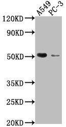
Western Blot Positive WB detected in: A549 whole cell lysate, PC-3 whole cell lysate All lanes: TNFRSF1A antibody at 1:2000 Secondary Goat polyclonal to rabbit IgG at 1/50000 dilution Predicted band size: 51, 39, 26, 25 kDa Observed band size: 51 kDa
. Section was blocked with 10% normal goat serum 30min at RT. Then primary antibody (1% BSA) was incubated at 4°C overnight. The primary is detected by a biotinylated secondary antibody and visualized using an HRP conjugated SP system.")
.")
Western Blot Positive WB detected in: A549 whole cell lysate, PC-3 whole cell lysate All lanes: TNFRSF1A antibody at 1:2000 Secondary Goat polyclonal to rabbit IgG at 1/50000 dilution Predicted band size: 51, 39, 26, 25 kDa Observed band size: 51 kDa
TNFRSF1A Antibody
CSB-PA023977LA01HU
ApplicationsImmunoFluorescence, Western Blot, ELISA, ImmunoHistoChemistry
Product group Antibodies
ReactivityHuman
TargetTNFRSF1A
Overview
- SupplierCusabio
- Product NameTNFRSF1A Antibody
- Delivery Days Customer20
- ApplicationsImmunoFluorescence, Western Blot, ELISA, ImmunoHistoChemistry
- CertificationResearch Use Only
- ClonalityPolyclonal
- ConjugateUnconjugated
- Gene ID7132
- Target nameTNFRSF1A
- Target descriptionTNF receptor superfamily member 1A
- Target synonymsCD120a, FPF, TBP1, TNF-R, TNF-R-I, TNF-R55, TNFAR, TNFR1, TNFR55, TNFR60, p55, p55-R, p60, tumor necrosis factor receptor superfamily member 1A, TNF-R1, TNF-RI, TNFR-I, tumor necrosis factor binding protein 1, tumor necrosis factor receptor type 1, tumor necrosis factor-alpha receptor
- HostRabbit
- IsotypeIgG
- Protein IDP19438
- Protein NameTumor necrosis factor receptor superfamily member 1A
- Scientific DescriptionReceptor for TNFSF2/TNF-alpha and homotrimeric TNFSF1/lymphotoxin-alpha. The adapter molecule FADD recruits caspase-8 to the activated receptor. The resulting death-inducing signaling complex (DISC) performs caspase-8 proteolytic activation which initiates the subsequent cascade of caspases (aspartate-specific cysteine proteases) mediating apoptosis. Contributes to the induction of non-cytocidal TNF effects including anti-viral state and activation of the acid sphingomyelinase.
- ReactivityHuman
- Storage Instruction-20°C or -80°C
- UNSPSC41116161
Related products
Product group Antibodies
Anti-TNFRSF1A AntibodyA49067
ApplicationsWestern Blot, ELISA
ReactivityHuman
- SizePrice
Product group Antibodies

TNFR-S274 AntibodyABX026117
ApplicationsFlow Cytometry, Western Blot, ELISA
- SizePrice
Product group Antibodies

ApplicationsELISA
ReactivityHuman
TargetTNFRSF1A
- SizePrice

Product group Antibodies
Anti-TNF Receptor I/TNFRSF1A Antibody Picoband(r)A00294-3-CARRIER-FREE
ApplicationsFlow Cytometry, Western Blot, ELISA, ImmunoHistoChemistry
ReactivityHuman, Mouse, Rat
TargetTNFRSF1A
- SizePrice
Product group Antibodies

Goat anti-TNFRSF1A / TNFR1EB11397
ApplicationsWestern Blot, ELISA
ReactivityHuman, Porcine, Rat
TargetTNFRSF1A
- SizePrice
Product group Antibodies
ApplicationsWestern Blot, ImmunoHistoChemistry
ReactivityMouse, Porcine, Rat
TargetTNFRSF1A
- SizePrice
Product group Antibodies
References
TNFR1 Polyclonal AntibodyBS-2941R
ApplicationsFlow Cytometry, ImmunoFluorescence, Western Blot, ELISA, ImmunoCytoChemistry, ImmunoHistoChemistry, ImmunoHistoChemistry Paraffin
ReactivityBovine, Canine, Equine, Human, Mouse, Porcine, Rabbit, Rat
TargetTNFRSF1A
- SizePrice
Product group Antibodies
TNF Receptor I antibodyGTX102710
ApplicationsWestern Blot
ReactivityHuman
TargetTNFRSF1A
- SizePrice

Product group Antibodies
Anti-TNFRSF1A AntibodyHPA004102
ApplicationsWestern Blot, ImmunoHistoChemistry
ReactivityHuman
TargetTNFRSF1A
- SizePrice