


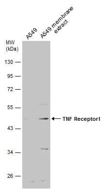
Figure 1. Western blot analysis of TNF Receptor I using anti-TNF Receptor I antibody (A00294-3). Electrophoresis was performed on a 5-20% SDS-PAGE gel at 70V (Stacking gel) / 90V (Resolving gel) for 2-3 hours. The sample well of each lane was loaded with 50ug of sample under reducing conditions. Lane 1: human Hela whole cell lysate, Lane 2: human K562 whole cell lysate, Lane 3: human Caco-2 whole cell lysate, Lane 4: rat liver tissue lysates, Lane 5: mouse small intestine tissue lysates, Lane 6: mouse liver tissue lysates, Lane 7: mouse HEPA1-6 whole cell lysate. After Electrophoresis, proteins were transferred to a Nitrocellulose membrane at 150mA for 50-90 minutes. Blocked the membrane with 5% Non-fat Milk/ TBS for 1.5 hour at RT. The membrane was incubated with rabbit anti-TNF Receptor I antigen affinity purified polyclonal antibody (Catalog # A00294-3) at 0.5 microg/mL overnight at 4°C, then washed with TBS-0.1%Tween 3 times with 5 minutes each and probed with a goat anti-rabbit IgG-HRP secondary antibody at a dilution of 1:10000 for 1.5 hour at RT. The signal is developed using an Enhanced Chemiluminescent detection (ECL) kit (Catalog # EK1002) with Tanon 5200 system. A specific band was detected for TNF Receptor I at approximately 50KD. The expected band size for TNF Receptor I is at 50KD.
. TNF Receptor I was detected in paraffin-embedded section of mouse kidney tissue. Heat mediated antigen retrieval was performed in citrate buffer (pH6, epitope retrieval solution) for 20 mins. The tissue section was blocked with 10% goat serum. The tissue section was then incubated with 1microg/ml rabbit anti-TNF Receptor I Antibody (A00294-3) overnight at 4°C. Biotinylated goat anti-rabbit IgG was used as secondary antibody and incubated for 30 minutes at 37°C. The tissue section was developed using Strepavidin-Biotin-Complex (SABC)(Catalog # SA1022) with DAB as the chromogen.")
. TNF Receptor I was detected in paraffin-embedded section of mouse liver tissue. Heat mediated antigen retrieval was performed in citrate buffer (pH6, epitope retrieval solution) for 20 mins. The tissue section was blocked with 10% goat serum. The tissue section was then incubated with 1microg/ml rabbit anti-TNF Receptor I Antibody (A00294-3) overnight at 4°C. Biotinylated goat anti-rabbit IgG was used as secondary antibody and incubated for 30 minutes at 37°C. The tissue section was developed using Strepavidin-Biotin-Complex (SABC)(Catalog # SA1022) with DAB as the chromogen.")
. TNF Receptor I was detected in paraffin-embedded section of mouse lung tissue. Heat mediated antigen retrieval was performed in citrate buffer (pH6, epitope retrieval solution) for 20 mins. The tissue section was blocked with 10% goat serum. The tissue section was then incubated with 1microg/ml rabbit anti-TNF Receptor I Antibody (A00294-3) overnight at 4°C. Biotinylated goat anti-rabbit IgG was used as secondary antibody and incubated for 30 minutes at 37°C. The tissue section was developed using Strepavidin-Biotin-Complex (SABC)(Catalog # SA1022) with DAB as the chromogen.")
. Overlay histogram showing U937 cells stained with A00294-3 (Blue line). To facilitate intracellular staining, cells were fixed with 4% paraformaldehyde and permeabilized with permeabilization buffer. The cells were blocked with 10% normal goat serum. And then incubated with rabbit anti-TNF Receptor I Antibody (A00294-3,1microg/1x106 cells) for 30 min at 20°C. DyLight®488 conjugated goat anti-rabbit IgG (BA1127, 5-10microg/1x106 cells) was used as secondary antibody for 30 minutes at 20°C. Isotype control antibody (Green line) was rabbit IgG (1microg/1x106) used under the same conditions. Unlabelled sample (Red line) was also used as a control.")
Figure 1. Western blot analysis of TNF Receptor I using anti-TNF Receptor I antibody (A00294-3). Electrophoresis was performed on a 5-20% SDS-PAGE gel at 70V (Stacking gel) / 90V (Resolving gel) for 2-3 hours. The sample well of each lane was loaded with 50ug of sample under reducing conditions. Lane 1: human Hela whole cell lysate, Lane 2: human K562 whole cell lysate, Lane 3: human Caco-2 whole cell lysate, Lane 4: rat liver tissue lysates, Lane 5: mouse small intestine tissue lysates, Lane 6: mouse liver tissue lysates, Lane 7: mouse HEPA1-6 whole cell lysate. After Electrophoresis, proteins were transferred to a Nitrocellulose membrane at 150mA for 50-90 minutes. Blocked the membrane with 5% Non-fat Milk/ TBS for 1.5 hour at RT. The membrane was incubated with rabbit anti-TNF Receptor I antigen affinity purified polyclonal antibody (Catalog # A00294-3) at 0.5 microg/mL overnight at 4°C, then washed with TBS-0.1%Tween 3 times with 5 minutes each and probed with a goat anti-rabbit IgG-HRP secondary antibody at a dilution of 1:10000 for 1.5 hour at RT. The signal is developed using an Enhanced Chemiluminescent detection (ECL) kit (Catalog # EK1002) with Tanon 5200 system. A specific band was detected for TNF Receptor I at approximately 50KD. The expected band size for TNF Receptor I is at 50KD.
Anti-TNF Receptor I/TNFRSF1A Antibody Picoband(r)
A00294-3-CARRIER-FREE
ApplicationsFlow Cytometry, Western Blot, ELISA, ImmunoHistoChemistry
Product group Antibodies
ReactivityHuman, Mouse, Rat
TargetTNFRSF1A
Overview
- SupplierBoster Bio
- Product NameAnti-TNF Receptor I/TNFRSF1A Antibody Picoband(r)
- Delivery Days Customer9
- Application Supplier NoteTested Species: In-house tested species with positive results. Predicted Species: Species predicted to be fit for the product based on sequence similarities. Other applications have not been tested. Optimal dilutions should be determined by end users.
- ApplicationsFlow Cytometry, Western Blot, ELISA, ImmunoHistoChemistry
- CertificationResearch Use Only
- ClonalityPolyclonal
- Concentration500 ug/ml
- Gene ID7132
- Target nameTNFRSF1A
- Target descriptionTNF receptor superfamily member 1A
- Target synonymsCD120a, FPF, TBP1, TNF-R, TNF-R-I, TNF-R55, TNFAR, TNFR1, TNFR55, TNFR60, p55, p55-R, p60, tumor necrosis factor receptor superfamily member 1A, TNF-R1, TNF-RI, TNFR-I, tumor necrosis factor binding protein 1, tumor necrosis factor receptor type 1, tumor necrosis factor-alpha receptor
- HostRabbit
- IsotypeIgG
- Protein IDP19438
- Protein NameTumor necrosis factor receptor superfamily member 1A
- Scientific DescriptionBoster Bio Anti-TNF Receptor I/TNFRSF1A Antibody Picoband® catalog # A00294-3. Tested in ELISA, IHC, WB applications. This antibody reacts with Human, Mouse, Rat. The brand Picoband indicates this is a premium antibody that guarantees superior quality, high affinity, and strong signals with minimal background in Western blot applications. Only our best-performing antibodies are designated as Picoband, ensuring unmatched performance.
- ReactivityHuman, Mouse, Rat
- Storage Instruction-20°C,2°C to 8°C
- UNSPSC12352203
Related products
Product group Antibodies
Anti-TNFRSF1A AntibodyA49067
ApplicationsWestern Blot, ELISA
ReactivityHuman
- SizePrice
Product group Antibodies

TNFR-S274 AntibodyABX026117
ApplicationsFlow Cytometry, Western Blot, ELISA
- SizePrice
Product group Antibodies

ApplicationsELISA
ReactivityHuman
TargetTNFRSF1A
- SizePrice
Product group Antibodies

Goat anti-TNFRSF1A / TNFR1EB11397
ApplicationsWestern Blot, ELISA
ReactivityHuman, Porcine, Rat
TargetTNFRSF1A
- SizePrice
Product group Antibodies
ApplicationsWestern Blot, ImmunoHistoChemistry
ReactivityMouse, Porcine, Rat
TargetTNFRSF1A
- SizePrice
Product group Antibodies
TNFRSF1A AntibodyCSB-PA023977LA01HU
ApplicationsImmunoFluorescence, Western Blot, ELISA, ImmunoHistoChemistry
ReactivityHuman
TargetTNFRSF1A
- SizePrice
Product group Antibodies
References
TNFR1 Polyclonal AntibodyBS-2941R
ApplicationsFlow Cytometry, ImmunoFluorescence, Western Blot, ELISA, ImmunoCytoChemistry, ImmunoHistoChemistry, ImmunoHistoChemistry Paraffin
ReactivityBovine, Canine, Equine, Human, Mouse, Porcine, Rabbit, Rat
TargetTNFRSF1A
- SizePrice
Product group Antibodies
TNF Receptor I antibodyGTX102710
ApplicationsWestern Blot
ReactivityHuman
TargetTNFRSF1A
- SizePrice

Product group Antibodies
Anti-TNFRSF1A AntibodyHPA004102
ApplicationsWestern Blot, ImmunoHistoChemistry
ReactivityHuman
TargetTNFRSF1A
- SizePrice