

IHC image of CSB-PA025468OA01HU diluted at 1:100 and staining in paraffin-embedded human colon cancer performed on a Leica BondTM system. After dewaxing and hydration, antigen retrieval was mediated by high pressure in a citrate buffer (pH 6.0). Section was blocked with 10% normal goat serum 30min at RT. Then primary antibody (1% BSA) was incubated at 4°C overnight. The primary is detected by a biotinylated secondary antibody and visualized using an HRP conjugated SP system.
. Section was blocked with 10% normal goat serum 30min at RT. Then primary antibody (1% BSA) was incubated at 4°C overnight. The primary is detected by a biotinylated secondary antibody and visualized using an HRP conjugated SP system.")
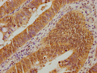
IHC image of CSB-PA025468OA01HU diluted at 1:100 and staining in paraffin-embedded human colon cancer performed on a Leica BondTM system. After dewaxing and hydration, antigen retrieval was mediated by high pressure in a citrate buffer (pH 6.0). Section was blocked with 10% normal goat serum 30min at RT. Then primary antibody (1% BSA) was incubated at 4°C overnight. The primary is detected by a biotinylated secondary antibody and visualized using an HRP conjugated SP system.
UBE2M Antibody
CSB-PA025468OA01HU
ApplicationsELISA, ImmunoHistoChemistry
Product group Antibodies
ReactivityHuman
TargetUBE2M
Overview
- SupplierCusabio
- Product NameUBE2M Antibody
- Delivery Days Customer20
- ApplicationsELISA, ImmunoHistoChemistry
- CertificationResearch Use Only
- ClonalityPolyclonal
- ConjugateUnconjugated
- Gene ID9040
- Target nameUBE2M
- Target descriptionubiquitin conjugating enzyme E2 M
- Target synonymsUBC-RS2, UBC12, hUbc12, NEDD8-conjugating enzyme Ubc12, NEDD8 carrier protein, NEDD8 protein ligase, epididymis secretory sperm binding protein, ubiquitin carrier protein M, ubiquitin conjugating enzyme E2M, ubiquitin-conjugating enzyme E2M (UBC12 homolog, yeast), ubiquitin-protein ligase M, yeast UBC12 homolog
- HostRabbit
- IsotypeIgG
- Protein IDP61081
- Protein NameNEDD8-conjugating enzyme Ubc12
- Scientific DescriptionAccepts the ubiquitin-like protein NEDD8 from the UBA3-NAE1 E1 complex and catalyzes its covalent attachment to other proteins. The specific interaction with the E3 ubiquitin ligase RBX1, but not RBX2, suggests that the RBX1-UBE2M complex neddylates specific target proteins, such as CUL1, CUL2, CUL3 and CUL4. Involved in cell proliferation.
- ReactivityHuman
- Storage Instruction-20°C or -80°C
- UNSPSC41116161
Related products
Product group Antibodies
UBE2M Polyclonal AntibodyBS-8382R
ApplicationsImmunoFluorescence, Western Blot, ELISA, ImmunoCytoChemistry, ImmunoHistoChemistry, ImmunoHistoChemistry Frozen, ImmunoHistoChemistry Paraffin
ReactivityBovine, Equine, Human, Mouse, Rat, Xenopus
TargetUBE2M
- SizePrice
Product group Antibodies

UBE2M / UBC12 AntibodyLS-C410542
ApplicationsWestern Blot
ReactivityHuman, Mouse
TargetUBE2M
- SizePrice
Product group Antibodies
UBE2M antibody [N1C2]GTX101653
ApplicationsWestern Blot, ImmunoHistoChemistry, ImmunoHistoChemistry Paraffin
ReactivityHuman
TargetUBE2M
- SizePrice

Product group Antibodies
Anti-UBE2M AntibodyHPA054551
ApplicationsWestern Blot, ImmunoCytoChemistry, ImmunoHistoChemistry
ReactivityHuman
TargetUBE2M
- SizePrice

Product group Antibodies
Anti-UBC12/UBE2M Antibody Picoband(r)A07478-1-CARRIER-FREE
ApplicationsFlow Cytometry, ImmunoFluorescence, Western Blot, ELISA, ImmunoCytoChemistry, ImmunoHistoChemistry
ReactivityHuman, Mouse, Rat
TargetUBE2M
- SizePrice