

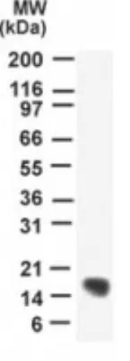
WB analysis of human heart tissue lysate using GTX13663 UBE2N antibody.
WB analysis of human heart tissue lysate using GTX13663 UBE2N antibody.
UBE2N antibody
GTX13663
ApplicationsImmunoPrecipitation, Western Blot
Product group Antibodies
ReactivityHuman
TargetUBE2N
Overview
- SupplierGeneTex
- Product NameUBE2N antibody
- Delivery Days Customer9
- Application Supplier NoteWB: 2 microg/ml. IP: 1:10 - 1:500. *Optimal dilutions/concentrations should be determined by the researcher.Not tested in other applications.
- ApplicationsImmunoPrecipitation, Western Blot
- CertificationResearch Use Only
- ClonalityPolyclonal
- Concentration0.5 mg/ml
- ConjugateUnconjugated
- Gene ID7334
- Target nameUBE2N
- Target descriptionubiquitin conjugating enzyme E2 N
- Target synonymsHEL-S-71, UBC13, UBCHBEN, UBCHBEN; UBC13, UbcH-ben, UbcH13, ubiquitin-conjugating enzyme E2 N, E2 ubiquitin-conjugating enzyme N, bendless-like ubiquitin conjugating enzyme, epididymis secretory protein Li 71, ubiquitin carrier protein N, ubiquitin conjugating enzyme E2N, ubiquitin-conjugating enzyme E2N (UBC13 homolog, yeast), ubiquitin-conjugating enzyme E2N (homologous to yeast UBC13), ubiquitin-protein ligase N, yeast UBC13 homolog
- HostRabbit
- IsotypeIgG
- Protein IDP61088
- Protein NameUbiquitin-conjugating enzyme E2 N
- Scientific DescriptionThe modification of proteins with ubiquitin is an important cellular mechanism for targeting abnormal or short-lived proteins for degradation. Ubiquitination involves at least three classes of enzymes: ubiquitin-activating enzymes, or E1s, ubiquitin-conjugating enzymes, or E2s, and ubiquitin-protein ligases, or E3s. This gene encodes a member of the E2 ubiquitin-conjugating enzyme family. Studies in mouse suggest that this protein plays a role in DNA postreplication repair. [provided by RefSeq]
- ReactivityHuman
- Storage Instruction-20°C or -80°C,2°C to 8°C
- UNSPSC41116161
Datasheet
Related products
Product group Antibodies
UBE2N AntibodyCSB-PA025470LA01HU
ApplicationsImmunoFluorescence, ELISA, ImmunoHistoChemistry
ReactivityHuman
TargetUBE2N
- SizePrice

Product group Antibodies
Anti-Ubc13/UBE2N Antibody Picoband(r)A02462-2-CARRIER-FREE
ApplicationsFlow Cytometry, ImmunoFluorescence, Western Blot, ELISA, ImmunoCytoChemistry
ReactivityHuman, Mouse, Rat
TargetUBE2N
- SizePrice
Product group Antibodies

UBE2N / UBC13 AntibodyLS-C832739
ApplicationsWestern Blot, ELISA, ImmunoHistoChemistry
ReactivityHuman, Mouse, Rat
TargetUBE2N
- SizePrice
Product group Antibodies
TargetUBE2N
- SizePrice

Product group Antibodies
Anti-UBE2N AntibodyHPA044976
ApplicationsWestern Blot, ImmunoCytoChemistry, ImmunoHistoChemistry
ReactivityHuman
TargetUBE2N
- SizePrice
Product group Antibodies
UBE2N Recombinant Antibody, AbBy Fluor-405 ConjugatedBSM-61921R-BF405
ApplicationsImmunoFluorescence, Western Blot
ReactivityHuman, Mouse, Rat
TargetUBE2N
- SizePrice
Product group Antibodies
UBE2N Polyclonal AntibodyCAC13238
ApplicationsImmunoFluorescence, ELISA, ImmunoHistoChemistry
TargetUBE2N
- SizePrice
Product group Antibodies
UBE2N antibodyGTX113290
ApplicationsImmunoFluorescence, Western Blot, ImmunoCytoChemistry
ReactivityHuman, Mouse
TargetUBE2N
- SizePrice
Product group Antibodies
UBE2N antibodyGTX48485
ApplicationsWestern Blot, ELISA
ReactivityHuman
TargetUBE2N
- SizePrice