

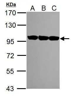
Various whole cell extracts (30 μg) were separated by 7.5% SDS-PAGE, and the membrane was blotted with VCP antibody [HL3018] (GTX640431) diluted at 1:1000. The HRP-conjugated anti-rabbit IgG antibody (GTX213110-01) was used to detect the primary antibody.
![Various tissue extracts (30 μg) were separated by 7.5% SDS-PAGE, and the membrane was blotted with VCP antibody [HL3018] (GTX640431) diluted at 1:1000. The HRP-conjugated anti-rabbit IgG antibody (GTX213110-01) was used to detect the primary antibody.](https://www.genetex.com/upload/website/prouct_img/normal/GTX640431/GTX640431_T-45425_20240531_WB_Z_tissue_24060619_963.webp "Various tissue extracts (30 μg) were separated by 7.5% SDS-PAGE, and the membrane was blotted with VCP antibody [HL3018] (GTX640431) diluted at 1:1000. The HRP-conjugated anti-rabbit IgG antibody (GTX213110-01) was used to detect the primary antibody.")
![Various whole cell extracts (30 μg) were separated by 7.5% SDS-PAGE, and the membrane was blotted with VCP antibody [HL3018] (GTX640431) diluted at 1:1000. The HRP-conjugated anti-rabbit IgG antibody (GTX213110-01) was used to detect the primary antibody.](https://www.genetex.com/upload/website/prouct_img/normal/GTX640431/GTX640431_T-45425_20240531_WB_M_24060619_647.webp "Various whole cell extracts (30 μg) were separated by 7.5% SDS-PAGE, and the membrane was blotted with VCP antibody [HL3018] (GTX640431) diluted at 1:1000. The HRP-conjugated anti-rabbit IgG antibody (GTX213110-01) was used to detect the primary antibody.")
![VCP antibody [HL3018] detects VCP protein by immunofluorescent analysis. Sample: HeLa cells were fixed in 4% paraformaldehyde at RT for 15 min. Green: VCP stained by VCP antibody [HL3018] (GTX640431) diluted at 1:500. Red: alpha Tubulin, a cytoskeleton marker, stained by alpha Tubulin antibody [GT114] (GTX628802) diluted at 1:1000. Blue: Fluoroshield with DAPI (GTX30920).](https://www.genetex.com/upload/website/prouct_img/normal/GTX640431/GTX640431_T-45425_20240621_ICC_IF_24070822_202.webp "VCP antibody [HL3018] detects VCP protein by immunofluorescent analysis. Sample: HeLa cells were fixed in 4% paraformaldehyde at RT for 15 min. Green: VCP stained by VCP antibody [HL3018] (GTX640431) diluted at 1:500. Red: alpha Tubulin, a cytoskeleton marker, stained by alpha Tubulin antibody [GT114] (GTX628802) diluted at 1:1000. Blue: Fluoroshield with DAPI (GTX30920).")
![VCP antibody [HL3018] detects VCP protein by immunohistochemical analysis. Sample: Paraffin-embedded human oral carcinoma. VCP stained by VCP antibody [HL3018] (GTX640431) diluted at 1:100. Antigen Retrieval: Citrate buffer, pH 6.0, 15 min](https://www.genetex.com/upload/website/prouct_img/normal/GTX640431/GTX640431_T-45425_20240719_IHC-P_24080622_168.webp "VCP antibody [HL3018] detects VCP protein by immunohistochemical analysis. Sample: Paraffin-embedded human oral carcinoma. VCP stained by VCP antibody [HL3018] (GTX640431) diluted at 1:100. Antigen Retrieval: Citrate buffer, pH 6.0, 15 min")
![VCP antibody [HL3018] detects VCP protein by immunohistochemical analysis. Sample: Paraffin-embedded mouse intestine. VCP stained by VCP antibody [HL3018] (GTX640431) diluted at 1:100. Antigen Retrieval: Citrate buffer, pH 6.0, 15 min](https://www.genetex.com/upload/website/prouct_img/normal/GTX640431/GTX640431_T-45425_20240719_IHC-P_M_24080622_660.webp "VCP antibody [HL3018] detects VCP protein by immunohistochemical analysis. Sample: Paraffin-embedded mouse intestine. VCP stained by VCP antibody [HL3018] (GTX640431) diluted at 1:100. Antigen Retrieval: Citrate buffer, pH 6.0, 15 min")
![VCP antibody [HL3018] detects VCP protein by immunohistochemical analysis. Sample: Paraffin-embedded rat brain. VCP stained by VCP antibody [HL3018] (GTX640431) diluted at 1:100. Antigen Retrieval: Citrate buffer, pH 6.0, 15 min](https://www.genetex.com/upload/website/prouct_img/normal/GTX640431/GTX640431_T-45425_20240719_IHC-P_R_24080622_473.webp "VCP antibody [HL3018] detects VCP protein by immunohistochemical analysis. Sample: Paraffin-embedded rat brain. VCP stained by VCP antibody [HL3018] (GTX640431) diluted at 1:100. Antigen Retrieval: Citrate buffer, pH 6.0, 15 min")
![Various whole cell extracts (30 μg) were separated by 7.5% SDS-PAGE, and the membrane was blotted with VCP antibody [HL3018] (GTX640431) diluted at 1:1000. The HRP-conjugated anti-rabbit IgG antibody (GTX213110-01) was used to detect the primary antibody.](https://www.genetex.com/upload/website/prouct_img/normal/GTX640431/GTX640431_45481_20240729_WB_24080602_640.webp "Various whole cell extracts (30 μg) were separated by 7.5% SDS-PAGE, and the membrane was blotted with VCP antibody [HL3018] (GTX640431) diluted at 1:1000. The HRP-conjugated anti-rabbit IgG antibody (GTX213110-01) was used to detect the primary antibody.")
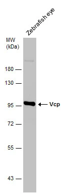
![Whole Japanese medaka extract (30 μg) was separated by 7.5% SDS-PAGE, and the membrane was blotted with VCP antibody [HL3018] (GTX640431) diluted at 1:1000. The HRP-conjugated anti-rabbit IgG antibody (GTX213110-01) was used to detect the primary antibody.](https://www.genetex.com/upload/website/prouct_img/normal/GTX640431/GTX640431_45481_20250815_WB_medaka_25082121_183.webp "Whole Japanese medaka extract (30 μg) was separated by 7.5% SDS-PAGE, and the membrane was blotted with VCP antibody [HL3018] (GTX640431) diluted at 1:1000. The HRP-conjugated anti-rabbit IgG antibody (GTX213110-01) was used to detect the primary antibody.")
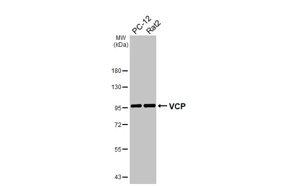
Various whole cell extracts (30 μg) were separated by 7.5% SDS-PAGE, and the membrane was blotted with VCP antibody [HL3018] (GTX640431) diluted at 1:1000. The HRP-conjugated anti-rabbit IgG antibody (GTX213110-01) was used to detect the primary antibody.
VCP antibody [HL3018]
GTX640431
ApplicationsImmunoFluorescence, Western Blot, ImmunoCytoChemistry, ImmunoHistoChemistry, ImmunoHistoChemistry Paraffin
Product group Antibodies
ReactivityHuman, Mouse, Rat, Zebra Fish
TargetVCP
Overview
- SupplierGeneTex
- Product NameVCP antibody [HL3018]
- Delivery Days Customer7
- Application Supplier NoteWB: 1:500-1:3000. *Optimal dilutions/concentrations should be determined by the researcher.Not tested in other applications.
- ApplicationsImmunoFluorescence, Western Blot, ImmunoCytoChemistry, ImmunoHistoChemistry, ImmunoHistoChemistry Paraffin
- CertificationResearch Use Only
- ClonalityMonoclonal
- Clone IDHL3018
- Concentration1 mg/ml
- ConjugateUnconjugated
- Gene ID7415
- Target nameVCP
- Target descriptionvalosin containing protein
- Target synonymsCDC48, FTDALS6, TERA, p97, transitional endoplasmic reticulum ATPase, 15S Mg(2+)-ATPase p97 subunit, TER ATPase
- HostRabbit
- IsotypeIgG
- Protein IDP55072
- Protein NameTransitional endoplasmic reticulum ATPase
- Scientific DescriptionThis gene encodes a member of the AAA ATPase family of proteins. The encoded protein plays a role in protein degradation, intracellular membrane fusion, DNA repair and replication, regulation of the cell cycle, and activation of the NF-kappa B pathway. This protein forms a homohexameric complex that interacts with a variety of cofactors and extracts ubiquitinated proteins from lipid membranes or protein complexes. Mutations in this gene cause IBMPFD (inclusion body myopathy with paget disease of bone and frontotemporal dementia), ALS (amyotrophic lateral sclerosis) and Charcot-Marie-Tooth disease in human patients. [provided by RefSeq, Aug 2017]
- ReactivityHuman, Mouse, Rat, Zebra Fish
- Storage Instruction-20°C or -80°C,2°C to 8°C
- UNSPSC12352203
Datasheet
Related products
Product group Antibodies
Anti-VCP Antibody144-60308
ApplicationsWestern Blot, ImmunoHistoChemistry
ReactivityHuman, Mouse, Rat
TargetVCP
- SizePrice

Product group Antibodies
Anti-VCP Antibody Picoband(r)A00610-2-CARRIER-FREE
ApplicationsFlow Cytometry, Western Blot, ELISA, ImmunoHistoChemistry
ReactivityHuman, Monkey, Mouse, Rat
TargetVCP
- SizePrice
Product group Antibodies
References
VCP antibodyGTX101089
ApplicationsImmunoPrecipitation, Western Blot, ImmunoHistoChemistry, ImmunoHistoChemistry Frozen, ImmunoHistoChemistry Paraffin
ReactivityHuman, Mouse, Rat, Zebra Fish
TargetVCP
- SizePrice
![Immunohistochemical analysis of paraffin-embedded zebrafish tissue, using VCP antibody [N1N2], N-term (GTX113030) at 1:300 dilution.](https://www.genetex.com/upload/website/prouct_img/normal/GTX113030/GTX113030_40079_IHC_Z_22111423_120.webp)
Product group Antibodies
References
VCP antibody [N1N2], N-termGTX113030
ApplicationsWestern Blot, ImmunoHistoChemistry, ImmunoHistoChemistry Paraffin
ReactivityCanine, Human, Mouse, Rat, Zebra Fish
TargetVCP
- SizePrice
Product group Antibodies
VCP antibody [N3C2], InternalGTX113099
ApplicationsWestern Blot, ImmunoHistoChemistry, ImmunoHistoChemistry Paraffin
ReactivityHuman, Mouse, Rat
TargetVCP
- SizePrice
Product group Antibodies
VCP Polyclonal AntibodyCAC15070
ApplicationsWestern Blot, ELISA, ImmunoHistoChemistry
TargetVCP
- SizePrice
Product group Antibodies
VCP Recombinant Antibody, AbBy Fluor-350 ConjugatedBSM-61451R-BF350
ApplicationsFlow Cytometry, ImmunoFluorescence, Western Blot, ImmunoCytoChemistry
ReactivityHuman, Mouse, Rat
TargetVCP
- SizePrice
Product group Antibodies
Anti-VCP AntibodyA98420
ApplicationsWestern Blot, ELISA
ReactivityHuman, Mouse, Rat
- SizePrice