

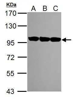
Sample (30 μg of whole cell lysate) A: NIH-3T3 B: JC C: BCL-1 7.5% SDS PAGE GTX113099 diluted at 1:1000 The HRP-conjugated anti-rabbit IgG antibody (GTX213110-01) was used to detect the primary antibody.
![VCP antibody [N3C2], Internal detects VCP protein at nucleus on human gastric cancer by immunohistochemical analysis. Sample: Paraffin-embedded gastric cancer. VCP antibody [N3C2], Internal (GTX113099) dilution: 1:500.
Antigen Retrieval: Trilogy? (EDTA based, pH 8.0) buffer, 15min](https://www.genetex.com/upload/website/prouct_img/normal/GTX113099/GTX113099_40457_IHC_w_23060500_723.webp "VCP antibody [N3C2], Internal detects VCP protein at nucleus on human gastric cancer by immunohistochemical analysis. Sample: Paraffin-embedded gastric cancer. VCP antibody [N3C2], Internal (GTX113099) dilution: 1:500.
Antigen Retrieval: Trilogy? (EDTA based, pH 8.0) buffer, 15min")
 were separated by 7.5% SDS-PAGE, and the membrane was blotted with VCP antibody (GTX113099) diluted by 1:1000.")
![VCP antibody [N3C2], Internal detects VCP protein by western blot analysis. A. 50 μg Rat brain lysate/extract 7.5% SDS-PAGE VCP antibody [N3C2], Internal (GTX113099) dilution: 1:10000 The HRP-conjugated anti-rabbit IgG antibody (GTX213110-01) was used to detect the primary antibody.](https://www.genetex.com/upload/website/prouct_img/normal/GTX113099/GTX113099_40457_WB_R_brain_w_23060500_728.webp "VCP antibody [N3C2], Internal detects VCP protein by western blot analysis. A. 50 μg Rat brain lysate/extract 7.5% SDS-PAGE VCP antibody [N3C2], Internal (GTX113099) dilution: 1:10000 The HRP-conjugated anti-rabbit IgG antibody (GTX213110-01) was used to detect the primary antibody.")
Sample (30 μg of whole cell lysate) A: NIH-3T3 B: JC C: BCL-1 7.5% SDS PAGE GTX113099 diluted at 1:1000 The HRP-conjugated anti-rabbit IgG antibody (GTX213110-01) was used to detect the primary antibody.
VCP antibody [N3C2], Internal
GTX113099
ApplicationsWestern Blot, ImmunoHistoChemistry, ImmunoHistoChemistry Paraffin
Product group Antibodies
ReactivityHuman, Mouse, Rat
TargetVCP
Overview
- SupplierGeneTex
- Product NameVCP antibody [N3C2], Internal
- Delivery Days Customer9
- Application Supplier NoteWB: 1:500-1:20000. IHC-P: 1:100-1:1000. *Optimal dilutions/concentrations should be determined by the researcher.Not tested in other applications.
- ApplicationsWestern Blot, ImmunoHistoChemistry, ImmunoHistoChemistry Paraffin
- CertificationResearch Use Only
- ClonalityPolyclonal
- Concentration0.2 mg/ml
- ConjugateUnconjugated
- Gene ID7415
- Target nameVCP
- Target descriptionvalosin containing protein
- Target synonymsCDC48, FTDALS6, TERA, p97, transitional endoplasmic reticulum ATPase, 15S Mg(2+)-ATPase p97 subunit, TER ATPase
- HostRabbit
- IsotypeIgG
- Protein IDP55072
- Protein NameTransitional endoplasmic reticulum ATPase
- Scientific DescriptionThe protein encoded by this gene is a member of a family that includes putative ATP-binding proteins involved in vesicle transport and fusion, 26S proteasome function, and assembly of peroxisomes. This protein, as a structural protein, is associated with clathrin, and heat-shock protein Hsc70, to form a complex. It has been implicated in a number of cellular events that are regulated during mitosis, including homotypic membrane fusion, spindle pole body function, and ubiquitin-dependent protein degradation. [provided by RefSeq]
- ReactivityHuman, Mouse, Rat
- Storage Instruction-20°C or -80°C,2°C to 8°C
- UNSPSC12352203
Datasheet
Related products
Product group Antibodies
Anti-VCP Antibody144-60308
ApplicationsWestern Blot, ImmunoHistoChemistry
ReactivityHuman, Mouse, Rat
TargetVCP
- SizePrice

Product group Antibodies
Anti-VCP Antibody Picoband(r)A00610-2-CARRIER-FREE
ApplicationsFlow Cytometry, Western Blot, ELISA, ImmunoHistoChemistry
ReactivityHuman, Monkey, Mouse, Rat
TargetVCP
- SizePrice
Product group Antibodies
References
VCP antibodyGTX101089
ApplicationsImmunoPrecipitation, Western Blot, ImmunoHistoChemistry, ImmunoHistoChemistry Frozen, ImmunoHistoChemistry Paraffin
ReactivityHuman, Mouse, Rat, Zebra Fish
TargetVCP
- SizePrice
![Immunohistochemical analysis of paraffin-embedded zebrafish tissue, using VCP antibody [N1N2], N-term (GTX113030) at 1:300 dilution.](https://www.genetex.com/upload/website/prouct_img/normal/GTX113030/GTX113030_40079_IHC_Z_22111423_120.webp)
Product group Antibodies
References
VCP antibody [N1N2], N-termGTX113030
ApplicationsWestern Blot, ImmunoHistoChemistry, ImmunoHistoChemistry Paraffin
ReactivityCanine, Human, Mouse, Rat, Zebra Fish
TargetVCP
- SizePrice
Product group Antibodies
VCP Polyclonal AntibodyCAC15070
ApplicationsWestern Blot, ELISA, ImmunoHistoChemistry
TargetVCP
- SizePrice
Product group Antibodies
VCP Recombinant Antibody, AbBy Fluor-350 ConjugatedBSM-61451R-BF350
ApplicationsFlow Cytometry, ImmunoFluorescence, Western Blot, ImmunoCytoChemistry
ReactivityHuman, Mouse, Rat
TargetVCP
- SizePrice
Product group Antibodies
Anti-VCP AntibodyA98420
ApplicationsWestern Blot, ELISA
ReactivityHuman, Mouse, Rat
- SizePrice
![Various whole cell extracts (30 μg) were separated by 7.5% SDS-PAGE, and the membrane was blotted with VCP antibody [HL3018] (GTX640431) diluted at 1:1000. The HRP-conjugated anti-rabbit IgG antibody (GTX213110-01) was used to detect the primary antibody.](https://www.genetex.com/upload/website/prouct_img/normal/GTX640431/GTX640431_T-45425_20240531_WB_R_24060619_377.webp)
Product group Antibodies
VCP antibody [HL3018]GTX640431
ApplicationsImmunoFluorescence, Western Blot, ImmunoCytoChemistry, ImmunoHistoChemistry, ImmunoHistoChemistry Paraffin
ReactivityHuman, Mouse, Rat, Zebra Fish
TargetVCP
- SizePrice