

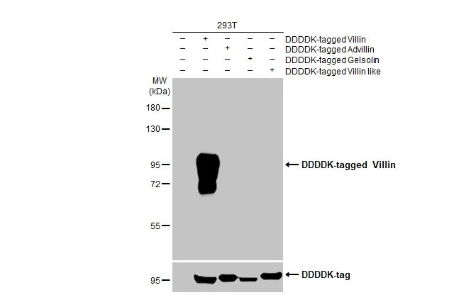
Non-transfected (–) and transfected (+) 293T whole cell extracts were separated by 7.5% SDS-PAGE, and the membrane was blotted with Villin antibody [HL1830] (GTX637555) diluted at 1:5000. The HRP-conjugated anti-rabbit IgG antibody (GTX213110-01) was used to detect the primary antibody.
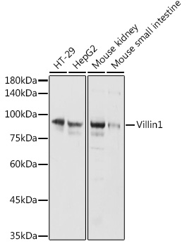
![Various whole cell extracts (30 μg) were separated by 7.5% SDS-PAGE, and the membrane was blotted with Villin antibody [HL1830] (GTX637555) diluted at 1:4000. The HRP-conjugated anti-rabbit IgG antibody (GTX213110-01) was used to detect the primary antibody. Corresponding RNA expression data for the same cell lines are based on Human Protein Atlas program.](https://www.genetex.com/upload/website/prouct_img/normal/GTX637555/GTX637555_44970_20230317_WB_TPM_watermark_23032022_712.webp "Various whole cell extracts (30 μg) were separated by 7.5% SDS-PAGE, and the membrane was blotted with Villin antibody [HL1830] (GTX637555) diluted at 1:4000. The HRP-conjugated anti-rabbit IgG antibody (GTX213110-01) was used to detect the primary antibody. Corresponding RNA expression data for the same cell lines are based on Human Protein Atlas program.")
![Whole cell extract (30 μg) was separated by 7.5% SDS-PAGE, and the membranes were blotted with Villin antibody [HL1830] (GTX637555) diluted at 1:4000 and competitor's antibody (#Highly competitor antibody) diluted at 1:1000. The HRP-conjugated anti-rabbit IgG antibody (GTX213110-01) was used to detect the primary antibody. *The competitor is not affiliated with GeneTex and does not endorse this product.](https://www.genetex.com/upload/website/prouct_img/normal/GTX637555/GTX637555_44970_20230707_WB_competitor_watermark_23071300_644.webp "Whole cell extract (30 μg) was separated by 7.5% SDS-PAGE, and the membranes were blotted with Villin antibody [HL1830] (GTX637555) diluted at 1:4000 and competitor's antibody (#Highly competitor antibody) diluted at 1:1000. The HRP-conjugated anti-rabbit IgG antibody (GTX213110-01) was used to detect the primary antibody. *The competitor is not affiliated with GeneTex and does not endorse this product.")
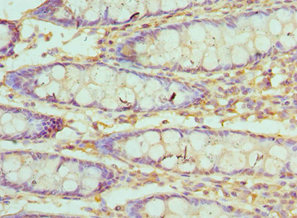
![Villin antibody [HL1830] detects Villin protein at cytoplasm by immunohistochemical analysis. Sample: Paraffin-embedded mouse duodenum. Villin stained by Villin antibody [HL1830] (GTX637555) diluted at 1:100. Antigen Retrieval: Citrate buffer, pH 6.0, 15 min](https://www.genetex.com/upload/website/prouct_img/normal/GTX637555/GTX637555_T-44823_20230714_IHC-P_M_23080901_576.webp "Villin antibody [HL1830] detects Villin protein at cytoplasm by immunohistochemical analysis. Sample: Paraffin-embedded mouse duodenum. Villin stained by Villin antibody [HL1830] (GTX637555) diluted at 1:100. Antigen Retrieval: Citrate buffer, pH 6.0, 15 min")
Non-transfected (–) and transfected (+) 293T whole cell extracts were separated by 7.5% SDS-PAGE, and the membrane was blotted with Villin antibody [HL1830] (GTX637555) diluted at 1:5000. The HRP-conjugated anti-rabbit IgG antibody (GTX213110-01) was used to detect the primary antibody.
Villin antibody [HL1830]
GTX637555
ApplicationsWestern Blot, ImmunoHistoChemistry, ImmunoHistoChemistry Paraffin
Product group Antibodies
ReactivityHuman, Mouse
TargetVIL1
Overview
- SupplierGeneTex
- Product NameVillin antibody [HL1830]
- Delivery Days Customer9
- ApplicationsWestern Blot, ImmunoHistoChemistry, ImmunoHistoChemistry Paraffin
- CertificationResearch Use Only
- ClonalityMonoclonal
- Clone IDHL1830
- Concentration0.5 mg/ml
- ConjugateUnconjugated
- Gene ID7429
- Target nameVIL1
- Target descriptionvillin 1
- Target synonymsD2S1471, VIL, villin-1
- HostRabbit
- IsotypeIgG
- Protein IDP09327
- Protein NameVillin-1
- Scientific DescriptionThis gene encodes a member of a family of calcium-regulated actin-binding proteins. This protein represents a dominant part of the brush border cytoskeleton which functions in the capping, severing, and bundling of actin filaments. Two mRNAs of 2.7 kb and 3.5 kb have been observed; they result from utilization of alternate poly-adenylation signals present in the terminal exon. [provided by RefSeq, Jul 2008]
- ReactivityHuman, Mouse
- Storage Instruction-20°C or -80°C,2°C to 8°C
- UNSPSC41116161
Datasheet
Related products
Product group Antibodies
VIL1 AntibodyCSB-PA025855ESR1HU
ApplicationsELISA, ImmunoHistoChemistry
ReactivityHuman
TargetVIL1
- SizePrice
Product group Antibodies
Villin (VIL) Polyclonal AntibodyCAU24074
ApplicationsWestern Blot, ImmunoHistoChemistry
ReactivityMouse, Porcine, Rat
TargetVIL1
- SizePrice
Product group Antibodies
Anti-Villin AntibodyA14804
ApplicationsWestern Blot
ReactivityHuman, Mouse, Rat
- SizePrice

Product group Antibodies
Anti-VIL1 AntibodyHPA006884
ApplicationsWestern Blot, ImmunoCytoChemistry, ImmunoHistoChemistry
ReactivityHuman
TargetVIL1
- SizePrice
Product group Antibodies

VIL1 / Villin AntibodyLS-C783283
ApplicationsWestern Blot
ReactivityHuman
TargetVIL1
- SizePrice

Product group Antibodies
Anti-Villin/VIL1 Antibody Picoband(r)PB9457-CARRIER-FREE
ApplicationsFlow Cytometry, ImmunoFluorescence, Western Blot, ImmunoHistoChemistry
ReactivityHamster, Human, Mouse, Rat
TargetVIL1
- SizePrice

Product group Antibodies
villin Recombinant AntibodyBSM-54212R
ApplicationsFlow Cytometry, ImmunoFluorescence, Western Blot, ImmunoCytoChemistry, ImmunoHistoChemistry, ImmunoHistoChemistry Paraffin
ReactivityHuman, Mouse, Rat
TargetVIL1
- SizePrice
![Whole cell extract (30 μg) was separated by 7.5% SDS-PAGE, and the membrane was blotted with Villin antibody [HL1756] (GTX637406) diluted at 1:5000. The HRP-conjugated anti-rabbit IgG antibody (GTX213110-01) was used to detect the primary antibody.](https://www.genetex.com/upload/website/prouct_img/normal/GTX637406/GTX637406_44956_20230224_WB_23030219_826.webp)
Product group Antibodies
Villin antibody [HL1756]GTX637406
ApplicationsWestern Blot
ReactivityHuman
TargetVIL1
- SizePrice
![IHC-P analysis of human renal cortex tissue using GTX04400 Villin antibody [MSVA-459R] HistoMAX?. A strong villin positivity is seen in proximal tubuli of the kidney but not in distal tubuli or glomeruli.](https://www.genetex.com/upload/website/prouct_img/normal/GTX04400/GTX04400_20230728_IHC-P_126_23072722_399.webp)
Product group Antibodies
ApplicationsImmunoHistoChemistry, ImmunoHistoChemistry Paraffin
ReactivityHuman
TargetVIL1
- SizePrice