

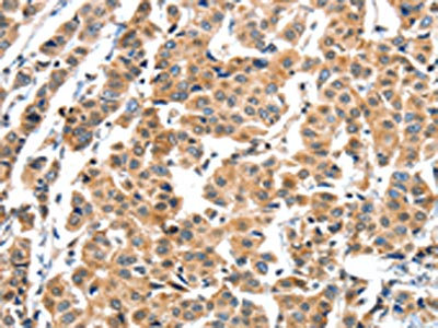
Immunohistochemistry of paraffin-embedded Human breast cancer tissue using XBP1 Polyclonal Antibody at dilution 1:60
Immunohistochemistry of paraffin-embedded Human breast cancer tissue using XBP1 Polyclonal Antibody at dilution 1:60
XBP1 Polyclonal Antibody
E-AB-10674
ApplicationsImmunoHistoChemistry
Product group Antibodies
TargetXBP1
Overview
- SupplierElabscience
- Product NameXBP1 Polyclonal Antibody
- Delivery Days Customer12
- ApplicationsImmunoHistoChemistry
- Applications SupplierELISA IHC
- CertificationResearch Use Only
- ClonalityPolyclonal
- Concentration0.2 mg/ml
- ConjugateUnconjugated
- Gene ID7494
- Target nameXBP1
- Target descriptionX-box binding protein 1
- Target synonymsTREB-5, TREB5, XBP-1, XBP2, X-box-binding protein 1, tax-responsive element-binding protein 5
- HostRabbit
- IsotypeIgG
- Protein IDP17861
- Protein NameX-box-binding protein 1
- Scientific DescriptionThis gene encodes a transcription factor that regulates MHC class II genes by binding to a promoter element referred to as an X box. This gene product is a bZIP protein, which was also identified as a cellular transcription factor that binds to an enhancer in the promoter of the T cell leukemia virus type 1 promoter. It may increase expression of viral proteins by acting as the DNA binding partner of a viral transactivator. It has been found that upon accumulation of unfolded proteins in the endoplasmic reticulum (ER), the mRNA of this gene is processed to an active form by an unconventional splicing mechanism that is mediated by the endonuclease inositol-requiring enzyme 1 (IRE1). The resulting loss of 26 nt from the spliced mRNA causes a frame-shift and an isoform XBP1(S), which is the functionally active transcription factor.
- Storage Instruction-20°C
- UNSPSC41116161
MSDS
Related products
Product group Antibodies
Anti-XBP1 AntibodyA13620
ApplicationsImmunoFluorescence, Western Blot, ImmunoCytoChemistry
ReactivityHuman, Mouse, Rat
- SizePrice
Product group Antibodies

Goat anti-XBP1 / TREB5EB08557
ApplicationsImmunoFluorescence, Western Blot, ELISA, ImmunoHistoChemistry
ReactivityCanine, Human, Mouse, Rat
TargetXBP1
- SizePrice
Product group Antibodies
XBP1 AntibodyCSB-PA027191
ApplicationsELISA, ImmunoHistoChemistry
ReactivityHuman
TargetXBP1
- SizePrice
Product group Antibodies

XBP1 AntibodyLS-C401118
ApplicationsELISA, ImmunoHistoChemistry
ReactivityHuman
TargetXBP1
- SizePrice

Product group Antibodies
Anti-XBP1 Antibody Picoband(r)PB9463-CARRIER-FREE
ApplicationsFlow Cytometry, ImmunoFluorescence, Western Blot, ImmunoCytoChemistry, ImmunoHistoChemistry
ReactivityHamster, Human, Mouse, Rat
TargetXBP1
- SizePrice
Product group Antibodies
References
XBP1 Polyclonal AntibodyBS-1668R
ApplicationsFlow Cytometry, ImmunoFluorescence, Western Blot, ELISA, ImmunoCytoChemistry, ImmunoHistoChemistry, ImmunoHistoChemistry Frozen, ImmunoHistoChemistry Paraffin
ReactivityBovine, Chicken, Human, Mouse, Rat
TargetXBP1
- SizePrice
![XBP1 antibody [N3C3] detects XBP1 protein at nucleus in mouse brain by immunohistochemical analysis. Sample: Paraffin-embedded mouse brain. XBP1 antibody [N3C3] (GTX102229) diluted at 1:500.
Antigen Retrieval: Citrate buffer, pH 6.0, 15 min](https://www.genetex.com/upload/website/prouct_img/normal/GTX102229/GTX102229_40079_20151130_IHC-P_M_w_23060100_471.webp)
Product group Antibodies
XBP1 antibody [N3C3]GTX102229
ApplicationsImmunoFluorescence, Western Blot, ImmunoCytoChemistry, ImmunoHistoChemistry, ImmunoHistoChemistry Paraffin
ReactivityHuman, Mouse, Rat
TargetXBP1
- SizePrice
Product group Antibodies
Anti-XBP1 Antibody144-01731
ApplicationsImmunoFluorescence, Western Blot
ReactivityHuman, Mouse, Rat
TargetXBP1
- SizePrice