

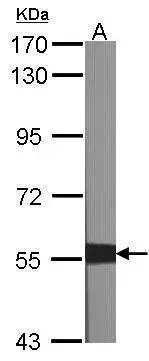
Sample (30 μg of whole cell lysate) A:NIH-3T3 7.5% SDS PAGE GTX100496 diluted at 1:1000 The HRP-conjugated anti-rabbit IgG antibody (GTX213110-01) was used to detect the primary antibody.
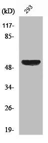
![XIAP antibody [C1C3] detects XIAP protein by western blot analysis. A. 30 μg 293T whole cell lysate/extract B. 30 μg A431 whole cell lysate/extract C. 30 μg H1299 whole cell lysate/extract 7.5% SDS-PAGE XIAP antibody [C1C3] (GTX100496) dilution: 1:500 The HRP-conjugated anti-rabbit IgG antibody (GTX213110-01) was used to detect the primary antibody.](https://www.genetex.com/upload/website/prouct_img/normal/GTX100496/GTX100496_39918_WB_w_23060100_122.webp "XIAP antibody [C1C3] detects XIAP protein by western blot analysis. A. 30 μg 293T whole cell lysate/extract B. 30 μg A431 whole cell lysate/extract C. 30 μg H1299 whole cell lysate/extract 7.5% SDS-PAGE XIAP antibody [C1C3] (GTX100496) dilution: 1:500 The HRP-conjugated anti-rabbit IgG antibody (GTX213110-01) was used to detect the primary antibody.")
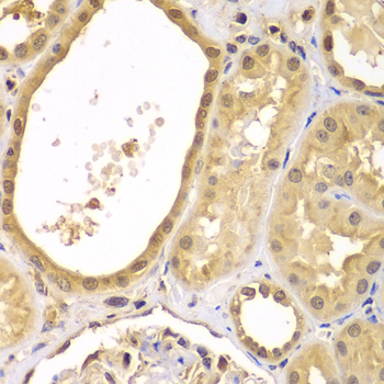
![XIAP antibody [C1C3] detects XIAP protein at cytoplasm in human breast carcinoma by immunohistochemical analysis. Sample: Paraffin-embedded human breast carcinoma. XIAP antibody [C1C3] (GTX100496) diluted at 1:500.
Antigen Retrieval: Trilogy? (EDTA based, pH 8.0) buffer, 15min](https://www.genetex.com/upload/website/prouct_img/normal/GTX100496/GTX100496_39918_20160125_IHC-P_w_23060100_944.webp "XIAP antibody [C1C3] detects XIAP protein at cytoplasm in human breast carcinoma by immunohistochemical analysis. Sample: Paraffin-embedded human breast carcinoma. XIAP antibody [C1C3] (GTX100496) diluted at 1:500.
Antigen Retrieval: Trilogy? (EDTA based, pH 8.0) buffer, 15min")
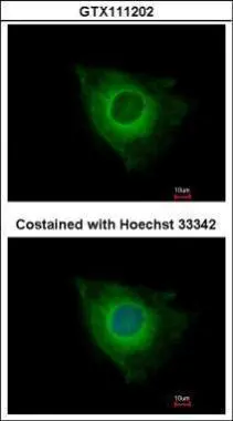
![XIAP antibody [C1C3] detects XIAP protein at cytoplasm by immunofluorescent analysis. Sample: HeLa cells were fixed in ice-cold MeOH for 5 min. Green: XIAP protein stained by XIAP antibody [C1C3] (GTX100496) diluted at 1:500. Blue: Hoechst 33342 staining. Scale bar = 10 μm.](https://www.genetex.com/upload/website/prouct_img/normal/GTX100496/GTX100496_39918_20150506_IFA_w_23060100_528.webp "XIAP antibody [C1C3] detects XIAP protein at cytoplasm by immunofluorescent analysis. Sample: HeLa cells were fixed in ice-cold MeOH for 5 min. Green: XIAP protein stained by XIAP antibody [C1C3] (GTX100496) diluted at 1:500. Blue: Hoechst 33342 staining. Scale bar = 10 μm.")
 antibody at 1:500 dilution.
Antigen Retrieval: Trilogy? (EDTA based, pH 8.0) buffer, 15min")
![XIAP antibody [C1C3] detects XIAP protein at cytoplasm on mouse spleen by immunohistochemical analysis. Sample: Paraffin-embedded mouse spleen. XIAP antibody [C1C3] (GTX100496) diluted at 1:500.
Antigen Retrieval: Trilogy? (EDTA based, pH 8.0) buffer, 15min](https://www.genetex.com/upload/website/prouct_img/normal/GTX100496/GTX100496_39918_20150206_IHC_M_w_23060100_534.webp "XIAP antibody [C1C3] detects XIAP protein at cytoplasm on mouse spleen by immunohistochemical analysis. Sample: Paraffin-embedded mouse spleen. XIAP antibody [C1C3] (GTX100496) diluted at 1:500.
Antigen Retrieval: Trilogy? (EDTA based, pH 8.0) buffer, 15min")
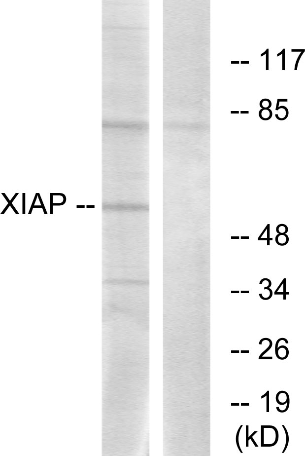
![Wild-type (WT) and XIAP knockout (KO) HeLa cell extracts (30 μg) were separated by 7.5% SDS-PAGE, and the membrane was blotted with XIAP antibody [C1C3] (GTX100496) diluted at 1:500. The HRP-conjugated anti-rabbit IgG antibody (GTX213110-01) was used to detect the primary antibody.](https://www.genetex.com/upload/website/prouct_img/normal/GTX100496/GTX100496_40205_20190322_WB_KO_watermark_24062501_515.webp "Wild-type (WT) and XIAP knockout (KO) HeLa cell extracts (30 μg) were separated by 7.5% SDS-PAGE, and the membrane was blotted with XIAP antibody [C1C3] (GTX100496) diluted at 1:500. The HRP-conjugated anti-rabbit IgG antibody (GTX213110-01) was used to detect the primary antibody.")
Sample (30 μg of whole cell lysate) A:NIH-3T3 7.5% SDS PAGE GTX100496 diluted at 1:1000 The HRP-conjugated anti-rabbit IgG antibody (GTX213110-01) was used to detect the primary antibody.
XIAP antibody [C1C3]
GTX100496
ApplicationsImmunoFluorescence, Western Blot, ImmunoCytoChemistry, ImmunoHistoChemistry, ImmunoHistoChemistry Paraffin
Product group Antibodies
ReactivityHuman, Mouse
TargetXIAP
Overview
- SupplierGeneTex
- Product NameXIAP antibody [C1C3]
- Delivery Days Customer9
- Application Supplier NoteWB: 1:500-1:3000. ICC/IF: 1:100-1:1000. IHC-P: 1:100-1:1000. *Optimal dilutions/concentrations should be determined by the researcher.Not tested in other applications.
- ApplicationsImmunoFluorescence, Western Blot, ImmunoCytoChemistry, ImmunoHistoChemistry, ImmunoHistoChemistry Paraffin
- CertificationResearch Use Only
- ClonalityPolyclonal
- Concentration1 mg/ml
- ConjugateUnconjugated
- Gene ID331
- Target nameXIAP
- Target descriptionX-linked inhibitor of apoptosis
- Target synonymsAPI3, BIRC4, IAP-3, ILP1, MIHA, XLP2, hIAP-3, hIAP3, E3 ubiquitin-protein ligase XIAP, IAP-like protein 1, RING-type E3 ubiquitin transferase XIAP, X-linked IAP, X-linked inhibitor of apoptosis, E3 ubiquitin protein ligase, baculoviral IAP repeat-containing protein 4, inhibitor of apoptosis protein 3
- HostRabbit
- IsotypeIgG
- Protein IDP98170
- Protein NameE3 ubiquitin-protein ligase XIAP
- Scientific DescriptionThe protein encoded by this gene is a member of a family of proteins which inhibit apoptosis through binding to tumor necrosis factor receptor-associated factors TRAF1 and TRAF2. This protein inhibits apoptosis induced by menadione, a potent inducer of free radicals, and ICE. It also inhibits at least two members of the caspase family of cell-death proteases, caspase-3 and caspase-7. [provided by RefSeq]
- ReactivityHuman, Mouse
- Storage Instruction-20°C or -80°C,2°C to 8°C
- UNSPSC41116161
Datasheet
Related products
Product group Antibodies
Anti-XIAP AntibodyA96107
ApplicationsWestern Blot, ELISA, ImmunoHistoChemistry
ReactivityHuman, Mouse, Rat
- SizePrice
Product group Antibodies
Anti-BIRC4 Antibody130-10444
ApplicationsWestern Blot, ELISA
TargetXIAP
- SizePrice
Product group Antibodies

ApplicationsWestern Blot, ELISA
ReactivityHuman
TargetXIAP
- SizePrice
Product group Antibodies
XIAP Polyclonal Antibodybs-55223R
ApplicationsImmunoFluorescence, Western Blot, ImmunoCytoChemistry, ImmunoHistoChemistry, ImmunoHistoChemistry Paraffin
ReactivityHuman, Mouse, Rat
TargetXIAP
- SizePrice
Product group Antibodies
ApplicationsImmunoFluorescence, Western Blot, ImmunoCytoChemistry, ImmunoHistoChemistry
ReactivityPorcine
TargetXIAP
- SizePrice
Product group Antibodies
XIAP AntibodyCSB-PA004519
ApplicationsWestern Blot, ELISA, ImmunoHistoChemistry
ReactivityHuman, Mouse, Rat
TargetXIAP
- SizePrice

Product group Antibodies
Anti-XIAP AntibodyHPA042428
ApplicationsImmunoHistoChemistry
ReactivityHuman
TargetXIAP
- SizePrice
Product group Antibodies
XIAP antibodyGTX111202
ApplicationsImmunoFluorescence, Western Blot, ImmunoCytoChemistry, ImmunoHistoChemistry, ImmunoHistoChemistry Paraffin
ReactivityHuman, Mouse
TargetXIAP
- SizePrice
![Wild-type (WT) and XIAP knockout (KO) HeLa cell extracts (30 μg) were separated by 7.5% SDS-PAGE, and the membrane was blotted with XIAP antibody [N1C1] (GTX113130) diluted at 1:500. The HRP-conjugated anti-rabbit IgG antibody (GTX213110-01) was used to detect the primary antibody.](https://www.genetex.com/upload/website/prouct_img/normal/GTX113130/GTX113130_40114_20170330_WB_KO_watermark_w_23060500_757.webp)
Product group Antibodies
XIAP antibody [N1C1]GTX113130
ApplicationsImmunoFluorescence, Western Blot, ImmunoCytoChemistry, ImmunoHistoChemistry, ImmunoHistoChemistry Paraffin
ReactivityHuman, Mouse
TargetXIAP
- SizePrice
Product group Antibodies
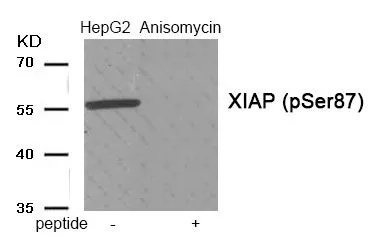
XIAP (phospho Ser87) antibodyGTX55394
ApplicationsWestern Blot
ReactivityHuman
TargetXIAP
- SizePrice