

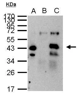
XPA antibody immunoprecipitates XPA protein in IP experiments. IP Sample: 1000 μg HeLa whole cell lysate/extract A. 40 μg HeLa whole cell lysate/extract B. Control with 2.5 μg of preimmune rabbit IgG C. Immunoprecipitation of XPA protein by 2.5 μg of XPA antibody (GTX103168) 12% SDS-PAGE The immunoprecipitated XPA protein was detected by XPA antibody (GTX103168) diluted at 1:1000. EasyBlot anti-rabbit IgG (GTX221666-01) was used as a secondary reagent.
 antibody at 1:200 dilution.")
 antibody at 1:500 dilution.
Antigen Retrieval: Trilogy? (EDTA based, pH 8.0) buffer, 15min")
 and transfected (+) 293T whole cell extracts (30 μg) were separated by 10% SDS-PAGE, and the membrane was blotted with XPA antibody (GTX103168) diluted at 1:2000.")
XPA antibody immunoprecipitates XPA protein in IP experiments. IP Sample: 1000 μg HeLa whole cell lysate/extract A. 40 μg HeLa whole cell lysate/extract B. Control with 2.5 μg of preimmune rabbit IgG C. Immunoprecipitation of XPA protein by 2.5 μg of XPA antibody (GTX103168) 12% SDS-PAGE The immunoprecipitated XPA protein was detected by XPA antibody (GTX103168) diluted at 1:1000. EasyBlot anti-rabbit IgG (GTX221666-01) was used as a secondary reagent.
XPA antibody
GTX103168
ApplicationsImmunoFluorescence, ImmunoPrecipitation, Western Blot, ImmunoCytoChemistry, ImmunoHistoChemistry, ImmunoHistoChemistry Paraffin
Product group Antibodies
ReactivityHuman, Mouse
TargetXPA
Overview
- SupplierGeneTex
- Product NameXPA antibody
- Delivery Days Customer9
- Application Supplier NoteWB: 1:500-1:3000. ICC/IF: 1:100-1:1000. IHC-P: 1:100-1:1000. IP: 1:500-1:1000. *Optimal dilutions/concentrations should be determined by the researcher.Not tested in other applications.
- ApplicationsImmunoFluorescence, ImmunoPrecipitation, Western Blot, ImmunoCytoChemistry, ImmunoHistoChemistry, ImmunoHistoChemistry Paraffin
- CertificationResearch Use Only
- ClonalityPolyclonal
- Concentration1 mg/ml
- ConjugateUnconjugated
- Gene ID7507
- Target nameXPA
- Target descriptionXPA, DNA damage recognition and repair factor
- Target synonymsXP1, XPAC, DNA repair protein complementing XP-A cells, xeroderma pigmentosum group A-complementing protein, xeroderma pigmentosum, complementation group A
- HostRabbit
- IsotypeIgG
- Protein IDP23025
- Protein NameDNA repair protein complementing XP-A cells
- Scientific DescriptionThis gene encodes a zinc finger protein involved in DNA excision repair. The encoded protein is part of the NER (nucleotide excision repair) complext which is responsible for repair of UV radiation-induced photoproducts and DNA adducts induced by chemical carcinogens. Mutations in this gene are associated with xeroderma pigmentosum complementation group A. Alternatively spliced transcript variants have been found for this gene. [provided by RefSeq]
- ReactivityHuman, Mouse
- Storage Instruction-20°C or -80°C,2°C to 8°C
- UNSPSC12352203
References
- Gautam A, Fawcett H, Burdova K, et al. APE1-dependent base excision repair of DNA photodimers in human cells. Mol Cell. 2023,83(20):3669-3678.e7. doi: 10.1016/j.molcel.2023.09.013Read this paper
- García-Carmona JA, Yousefzadeh MJ, Alarcón-Soldevilla F, et al. Case Report: Identification of a Heterozygous XPA c.553C>T Mutation Causing Neurological Impairment in a Case of Xeroderma Pigmentosum Complementation Group A. Front Genet. 2021,12:717361. doi: 10.3389/fgene.2021.717361Read this paper
- Chitale S, Richly H. DICER- and MMSET-catalyzed H4K20me2 recruits the nucleotide excision repair factor XPA to DNA damage sites. J Cell Biol. 2018,217(2):527-540. doi: 10.1083/jcb.201704028Read this paper
- Manandhar M, Lowery MG, Boulware KS, et al. Transcriptional consequences of XPA disruption in human cell lines. DNA Repair (Amst). 2017,57:76-90. doi: 10.1016/j.dnarep.2017.06.028Read this paper
- Chitale S, Richly H. DICER and ZRF1 contribute to chromatin decondensation during nucleotide excision repair. Nucleic Acids Res. 2017,45(10):5901-5912. doi: 10.1093/nar/gkx261Read this paper
Datasheet
Related products
Product group Antibodies
Anti-XPA Antibody144-01626
ApplicationsImmunoFluorescence, Western Blot
ReactivityHuman, Mouse
TargetXPA
- SizePrice

Product group Antibodies
Anti-XPA Antibody Picoband(r)A01182-4-CARRIER-FREE
ApplicationsFlow Cytometry, ImmunoFluorescence, Western Blot, ELISA, ImmunoCytoChemistry
ReactivityHuman
TargetXPA
- SizePrice
![ICC/IF analysis of GM0637 cells irradiated with UV at 20 J/m2 using GTX00704 XPA antibody [5F12]. Dilution : 1:100](https://www.genetex.com/upload/website/prouct_img/normal/GTX00704/GTX00704_20191104_ICC-IF_w_23053121_680.webp)
Product group Antibodies
References
XPA antibody [5F12]GTX00704
ApplicationsImmunoFluorescence, Western Blot, ELISA, ImmunoCytoChemistry, Neutralisation/Blocking
ReactivityHuman
TargetXPA
- SizePrice
Product group Antibodies
ApplicationsImmunoPrecipitation, Western Blot, ImmunoCytoChemistry, ImmunoHistoChemistry
TargetXPA
- SizePrice
Product group Antibodies
Anti-XPA AntibodyA98418
ApplicationsWestern Blot, ELISA, ImmunoHistoChemistry
ReactivityHuman, Mouse
- SizePrice
Product group Antibodies
XPA antibodyGTX10905
ApplicationsWestern Blot, ImmunoHistoChemistry, ImmunoHistoChemistry Paraffin
ReactivityHuman
TargetXPA
- SizePrice
Product group Antibodies
XPA antibodyGTX20044
ApplicationsDot Blot, ImmunoFluorescence, Western Blot, ELISA, ImmunoCytoChemistry, Other Application
ReactivityHuman
TargetXPA
- SizePrice
Product group Antibodies
XPA AntibodyCSB-PA004520
ApplicationsWestern Blot, ELISA, ImmunoHistoChemistry
ReactivityHuman, Mouse
TargetXPA
- SizePrice
Product group Antibodies

XPA AntibodyLS-C331594
ApplicationsImmunoFluorescence, Western Blot, ImmunoHistoChemistry
ReactivityHuman, Mouse
TargetXPA
- SizePrice

Product group Antibodies
Anti-XPA AntibodyHPA056856
ApplicationsImmunoCytoChemistry
ReactivityHuman
TargetXPA
- SizePrice