

XPC antibody
GTX70309
ApplicationsImmunoFluorescence, ImmunoPrecipitation, Western Blot, ImmunoCytoChemistry
Product group Antibodies
ReactivityHuman
TargetXPC
Overview
- SupplierGeneTex
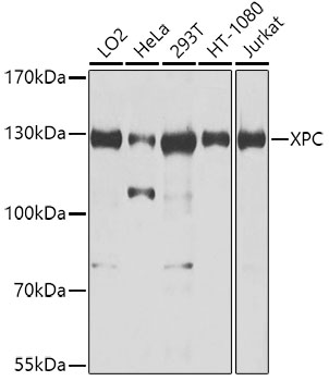
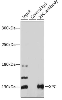
- Product NameXPC antibody
- Delivery Days Customer9
- Application Supplier NoteOptimal dilutions/concentrations should be determined by the researcher.
- ApplicationsImmunoFluorescence, ImmunoPrecipitation, Western Blot, ImmunoCytoChemistry
- CertificationResearch Use Only
- ClonalityPolyclonal
- Concentration0.5 mg/ml
- ConjugateUnconjugated
- Gene ID7508
- Target nameXPC
- Target descriptionXPC complex subunit, DNA damage recognition and repair factor
- Target synonymsRAD4, XP3, XPCC, p125, DNA repair protein complementing XP-C cells, mutant xeroderma pigmentosum group C, xeroderma pigmentosum, complementation group C
- HostRabbit
- IsotypeIgG
- Protein IDQ01831
- Protein NameDNA repair protein complementing XP-C cells
- Scientific DescriptionThe protein encoded by this gene is a key component of the XPC complex, which plays an important role in the early steps of global genome nucleotide excision repair (NER). The encoded protein is important for damage sensing and DNA binding, and shows a preference for single-stranded DNA. Mutations in this gene or some other NER components can result in Xeroderma pigmentosum, a rare autosomal recessive disorder characterized by increased sensitivity to sunlight with the development of carcinomas at an early age. Alternatively spliced transcript variants have been found for this gene. [provided by RefSeq, Aug 2017]
- ReactivityHuman
- Storage Instruction-20°C or -80°C,2°C to 8°C
- UNSPSC12352203
References
- Robu M, Shah RG, Shah GM. Methods to Study Intracellular Movement and Localization of the Nucleotide Excision Repair Proteins at the DNA Lesions in Mammalian Cells. Front Cell Dev Biol. 2020,8:590242. doi: 10.3389/fcell.2020.590242Read this paper
- Robu M, Shah RG, Petitclerc N, et al. Role of poly(ADP-ribose) polymerase-1 in the removal of UV-induced DNA lesions by nucleotide excision repair. Proc Natl Acad Sci U S A. 2013,110(5):1658-63. doi: 10.1073/pnas.1209507110Read this paper
- Muniandy PA, Thapa D, Thazhathveetil AK, et al. Repair of laser-localized DNA interstrand cross-links in G1 phase mammalian cells. J Biol Chem. 2009,284(41):27908-27917. doi: 10.1074/jbc.M109.029025Read this paper
- Liu Y, Wang Y, Rusinol AE, et al. Involvement of xeroderma pigmentosum group A (XPA) in progeria arising from defective maturation of prelamin A. FASEB J. 2008,22(2):603-11.Read this paper
Datasheet
Related products
Product group Antibodies
Anti-XPC AntibodyA10400
ApplicationsImmunoPrecipitation, Western Blot, ImmunoHistoChemistry
ReactivityHuman
- SizePrice
Product group Antibodies
XPC Polyclonal AntibodyBS-25269R
ApplicationsWestern Blot, ELISA
ReactivityBovine, Equine, Human, Mouse, Porcine, Rat
TargetXPC
- SizePrice
Product group Antibodies
References
XPC antibody [3.26]GTX70294
ApplicationsImmunoFluorescence, Western Blot, ImmunoCytoChemistry, ImmunoHistoChemistry, ImmunoHistoChemistry Paraffin
ReactivityHuman, Mouse
TargetXPC
- SizePrice
Product group Antibodies
References
XPC antibodyGTX70308
ApplicationsWestern Blot
ReactivityHuman
TargetXPC
- SizePrice
Product group Antibodies
Anti-XPC Antibody144-08354
ApplicationsImmunoPrecipitation, Western Blot, ImmunoHistoChemistry
ReactivityHuman
TargetXPC
- SizePrice
Product group Antibodies
XPC antibodyGTX55845
ApplicationsImmunoPrecipitation, Western Blot, ImmunoHistoChemistry, ImmunoHistoChemistry Paraffin
ReactivityHuman
TargetXPC
- SizePrice
Product group Antibodies
XPC antibody [HL2892]GTX640229
ApplicationsImmunoFluorescence, Western Blot, ImmunoCytoChemistry
ReactivityHuman
TargetXPC
- SizePrice
Product group Antibodies
XPC antibody [HL2894]GTX640231
ApplicationsWestern Blot, ImmunoHistoChemistry, ImmunoHistoChemistry Paraffin
ReactivityHuman, Mouse, Rat
TargetXPC
- SizePrice
Product group Antibodies
XPC antibody [C2C3], C-termGTX102840
ApplicationsWestern Blot, ImmunoHistoChemistry, ImmunoHistoChemistry Paraffin
ReactivityHuman
TargetXPC
- SizePrice