

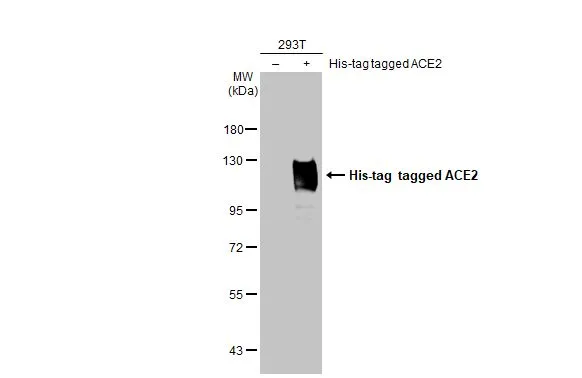
Non-transfected (–) and transfected (+) 293T whole cell extracts (30 μg) were separated by 7.5% SDS-PAGE, and the membrane was blotted with ACE2 antibody [HL1092] (GTX636265) diluted at 1:5000. The HRP-conjugated anti-rabbit IgG antibody (GTX213110-01) was used to detect the primary antibody.
![ACE2 antibody [HL1092] detects ACE2 protein at cell membrane by immunohistochemical analysis. Sample: Paraffin-embedded cat colon. ACE2 stained by ACE2 antibody [HL1092] (GTX636265) diluted at 1:100. Antigen Retrieval: Citrate buffer, pH 6.0, 15 min](https://www.genetex.com/upload/website/prouct_img/normal/GTX636265/GTX636265_44389_20230203_IHC-P_Cat_23020621_546.webp "ACE2 antibody [HL1092] detects ACE2 protein at cell membrane by immunohistochemical analysis. Sample: Paraffin-embedded cat colon. ACE2 stained by ACE2 antibody [HL1092] (GTX636265) diluted at 1:100. Antigen Retrieval: Citrate buffer, pH 6.0, 15 min")
![ACE2 antibody [HL1092] detects ACE2 protein at cell membrane by immunohistochemical analysis. Sample: Paraffin-embedded dog stomach. ACE2 stained by ACE2 antibody [HL1092] (GTX636265) diluted at 1:100. Antigen Retrieval: Citrate buffer, pH 6.0, 15 min](https://www.genetex.com/upload/website/prouct_img/normal/GTX636265/GTX636265_44389_20230203_IHC-P_Dog_23020621_640.webp "ACE2 antibody [HL1092] detects ACE2 protein at cell membrane by immunohistochemical analysis. Sample: Paraffin-embedded dog stomach. ACE2 stained by ACE2 antibody [HL1092] (GTX636265) diluted at 1:100. Antigen Retrieval: Citrate buffer, pH 6.0, 15 min")
![ACE2 antibody [HL1092] detects ACE2 protein at cell membrane by immunofluorescent analysis. Sample: VeroE6 cells were fixed in ice-cold MeOH for 5 min. Green: ACE2 stained by ACE2 antibody [HL1092] (GTX636265) diluted at 1:500. Red: alpha Tubulin, a cytoskeleton marker, stained by alpha Tubulin antibody [GT114] (GTX628802) diluted at 1:1000. Blue: Fluoroshield with DAPI (GTX30920).](https://www.genetex.com/upload/website/prouct_img/normal/GTX636265/GTX636265_44389_20220325_ICC_IF_w_23061202_819.webp "ACE2 antibody [HL1092] detects ACE2 protein at cell membrane by immunofluorescent analysis. Sample: VeroE6 cells were fixed in ice-cold MeOH for 5 min. Green: ACE2 stained by ACE2 antibody [HL1092] (GTX636265) diluted at 1:500. Red: alpha Tubulin, a cytoskeleton marker, stained by alpha Tubulin antibody [GT114] (GTX628802) diluted at 1:1000. Blue: Fluoroshield with DAPI (GTX30920).")
![Various whole cell and tissue extracts were separated by 7.5% SDS-PAGE, and the membrane was blotted with ACE2 antibody [HL1092] (GTX636265) diluted at 1:1000. The HRP-conjugated anti-rabbit IgG antibody (GTX213110-01) was used to detect the primary antibody, and the signal was developed with Trident femto Western HRP Substrate.](https://www.genetex.com/upload/website/prouct_img/normal/GTX636265/GTX636265_44389_20210806_WB_H_M_kidney_w_23061202_811.webp "Various whole cell and tissue extracts were separated by 7.5% SDS-PAGE, and the membrane was blotted with ACE2 antibody [HL1092] (GTX636265) diluted at 1:1000. The HRP-conjugated anti-rabbit IgG antibody (GTX213110-01) was used to detect the primary antibody, and the signal was developed with Trident femto Western HRP Substrate.")
![Non-transfected (–) and transfected (+) 293T whole cell extracts (50 μg) were separated by 5% SDS-PAGE, and the membrane was blotted with ACE2 antibody [HL1092] (GTX636265) diluted at 1:1000. The HRP-conjugated anti-rabbit IgG antibody (GTX213110-01) was used to detect the primary antibody, and the signal was developed with Trident ECL plus-Enhanced.](https://www.genetex.com/upload/website/prouct_img/normal/GTX636265/GTX636265_44389_20210806_WB_shRNA_watermark_w_23061202_559.webp "Non-transfected (–) and transfected (+) 293T whole cell extracts (50 μg) were separated by 5% SDS-PAGE, and the membrane was blotted with ACE2 antibody [HL1092] (GTX636265) diluted at 1:1000. The HRP-conjugated anti-rabbit IgG antibody (GTX213110-01) was used to detect the primary antibody, and the signal was developed with Trident ECL plus-Enhanced.")
![Various whole cell extracts (30 μg) were separated by 7.5% SDS-PAGE, and the membrane was blotted with ACE2 antibody [HL1092] (GTX636265) diluted at 1:1000. The HRP-conjugated anti-rabbit IgG antibody (GTX213110-01) was used to detect the primary antibody.](https://www.genetex.com/upload/website/prouct_img/normal/GTX636265/GTX636265_44389_20220506_WB_Monkey_H_w_23061202_677.webp "Various whole cell extracts (30 μg) were separated by 7.5% SDS-PAGE, and the membrane was blotted with ACE2 antibody [HL1092] (GTX636265) diluted at 1:1000. The HRP-conjugated anti-rabbit IgG antibody (GTX213110-01) was used to detect the primary antibody.")
![Untreated (–) and treated (+) VeroE6 whole cell extract (30 μg) were separated by 7.5% SDS-PAGE, and the membrane was blotted with ACE2 antibody [HL1092] (GTX636265) diluted at 1:1000. The HRP-conjugated anti-rabbit IgG antibody (GTX213110-01) was used to detect the primary antibody.](https://www.genetex.com/upload/website/prouct_img/normal/GTX636265/GTX636265_44907_20230707_WB_treatment_Tunicamycin_23081619_625.webp "Untreated (–) and treated (+) VeroE6 whole cell extract (30 μg) were separated by 7.5% SDS-PAGE, and the membrane was blotted with ACE2 antibody [HL1092] (GTX636265) diluted at 1:1000. The HRP-conjugated anti-rabbit IgG antibody (GTX213110-01) was used to detect the primary antibody.")
![Various whole cell extracts (30 μg) were separated by 5% SDS-PAGE, and the membrane was blotted with ACE2 antibody [HL1092] (GTX636265) diluted at 1:1000. The HRP-conjugated anti-rabbit IgG antibody (GTX213110-01) was used to detect the primary antibody, and the signal was developed with Trident femto Western HRP Substrate. Corresponding RNA expression data for the same cell lines are based on Human Protein Atlas program.](https://www.genetex.com/upload/website/prouct_img/normal/GTX636265/GTX636265_44907_20240119_WB_TPM_watermark_24012217_476.webp "Various whole cell extracts (30 μg) were separated by 5% SDS-PAGE, and the membrane was blotted with ACE2 antibody [HL1092] (GTX636265) diluted at 1:1000. The HRP-conjugated anti-rabbit IgG antibody (GTX213110-01) was used to detect the primary antibody, and the signal was developed with Trident femto Western HRP Substrate. Corresponding RNA expression data for the same cell lines are based on Human Protein Atlas program.")
![ACE2 antibody [HL1092] detects ACE2 protein by immunohistochemical analysis. Sample: Paraffin-embedded human tissues. ACE2 stained by ACE2 antibody [HL1092] (GTX636265) diluted at 1:10000. Antigen Retrieval: Tris-EDTA buffer, pH 9.0, 15 min](https://www.genetex.com/upload/website/prouct_img/normal/GTX636265/GTX636265_45278_20250808_IHC-P_Multiple_RPKM_2_25081423_173.webp "ACE2 antibody [HL1092] detects ACE2 protein by immunohistochemical analysis. Sample: Paraffin-embedded human tissues. ACE2 stained by ACE2 antibody [HL1092] (GTX636265) diluted at 1:10000. Antigen Retrieval: Tris-EDTA buffer, pH 9.0, 15 min")
Non-transfected (–) and transfected (+) 293T whole cell extracts (30 μg) were separated by 7.5% SDS-PAGE, and the membrane was blotted with ACE2 antibody [HL1092] (GTX636265) diluted at 1:5000. The HRP-conjugated anti-rabbit IgG antibody (GTX213110-01) was used to detect the primary antibody.
ACE2 antibody [HL1092]
GTX636265
ApplicationsImmunoFluorescence, Western Blot, ImmunoCytoChemistry, ImmunoHistoChemistry, ImmunoHistoChemistry Paraffin
Product group Antibodies
ReactivityCanine, Feline, Human, Monkey
TargetACE2
Overview
- SupplierGeneTex
- Product NameACE2 antibody [HL1092]
- Delivery Days Customer9
- ApplicationsImmunoFluorescence, Western Blot, ImmunoCytoChemistry, ImmunoHistoChemistry, ImmunoHistoChemistry Paraffin
- CertificationResearch Use Only
- ClonalityMonoclonal
- Clone IDHL1092
- Concentration1 mg/ml
- ConjugateUnconjugated
- Gene ID59272
- Target nameACE2
- Target descriptionangiotensin converting enzyme 2
- Target synonymsACEH, angiotensin-converting enzyme 2, ACE-related carboxypeptidase, angiotensin I converting enzyme (peptidyl-dipeptidase A) 2, angiotensin I converting enzyme 2, angiotensin-converting enzyme homolog, angiotensin-converting enzyme-related carboxypeptidase, metalloprotease MPROT15, peptidyl-dipeptidase A
- HostRabbit
- IsotypeIgG
- Protein IDQ9BYF1
- Protein NameAngiotensin-converting enzyme 2
- Scientific DescriptionThe protein encoded by this gene belongs to the angiotensin-converting enzyme family of dipeptidyl carboxydipeptidases and has considerable homology to human angiotensin 1 converting enzyme. This secreted protein catalyzes the cleavage of angiotensin I into angiotensin 1-9, and angiotensin II into the vasodilator angiotensin 1-7. The organ- and cell-specific expression of this gene suggests that it may play a role in the regulation of cardiovascular and renal function, as well as fertility. In addition, the encoded protein is a functional receptor for the spike glycoprotein of the human coronaviruses SARS and HCoV-NL63. [provided by RefSeq, Jul 2008]
- ReactivityCanine, Feline, Human, Monkey
- Storage Instruction-20°C or -80°C,2°C to 8°C
- UNSPSC12352203
Datasheet
Related products
Product group Antibodies

Anti-ACE2 [h11B11]AB03703-1.1
ApplicationsFlow Cytometry, ELISA, Neutralisation/Blocking
ReactivityHuman, Monkey
TargetACE2
- SizePrice

Product group Antibodies
Anti-ACE2 AntibodyAMAB91259
ApplicationsWestern Blot, ImmunoHistoChemistry
ReactivityHuman
TargetACE2
- SizePrice
Product group Antibodies
Anti-ACE2 Antibody130-10857
ApplicationsELISA
ReactivityVirus
TargetACE2
- SizePrice

Product group Antibodies
Anti-ACE2 Antibody Picoband(r)A00756-3-CARRIER-FREE
ApplicationsFlow Cytometry, Western Blot, ELISA
ReactivityHuman
TargetACE2
- SizePrice
![ACE2 antibody [SN0754] detects ACE2 protein at cell membrane by immunohistochemical analysis. Sample: Paraffin-embedded mouse kidney. ACE2 stained by ACE2 antibody [SN0754] (GTX01160) diluted at 1:2000. Antigen Retrieval: Citrate buffer, pH 6.0, 15 min](https://www.genetex.com/upload/website/prouct_img/normal/GTX01160/GTX01160_HK0921_20200313_IHC-P_M_w_23053121_166.webp)
Product group Antibodies
References
ACE2 antibody [SN0754]GTX01160
ApplicationsImmunoFluorescence, Western Blot, ImmunoCytoChemistry, ImmunoHistoChemistry, ImmunoHistoChemistry Paraffin
ReactivityHuman, Monkey, Mouse, Rat
TargetACE2
- SizePrice
![IHC-P analysis of human testis tissue using GTX04425 ACE2 antibody [MSVA-919R] HistoMAX?. Leydig cells and spermatocytes of the testis show a particularly strong ACE2 immunostaining in the testis.](https://www.genetex.com/upload/website/prouct_img/normal/GTX04425/GTX04425_20230728_IHC-P_2_23072722_840.webp)
Product group Antibodies
ApplicationsImmunoHistoChemistry, ImmunoHistoChemistry Paraffin
ReactivityHuman
TargetACE2
- SizePrice
![Mouse tissue extract (50 μg) was separated by 7.5% SDS-PAGE, and the membrane was blotted with ACE2 antibody [N1N2], N-term (GTX101395) diluted at 1:500. The HRP-conjugated anti-rabbit IgG antibody (GTX213110-01) was used to detect the primary antibody, and the signal was developed with Trident ECL plus-Enhanced.](https://www.genetex.com/upload/website/prouct_img/normal/GTX101395/GTX101395_43824_20200327_WB_M_kidney_w_23060100_326.webp)
Product group Antibodies
References
ACE2 antibody [N1N2], N-termGTX101395
ApplicationsFlow Cytometry, ImmunoFluorescence, Western Blot, ELISA, ImmunoCytoChemistry, ImmunoHistoChemistry, ImmunoHistoChemistry Paraffin
ReactivityHuman, Monkey, Mouse, Rat
TargetACE2
- SizePrice
![Whole cell extract (30 μg) was separated by 7.5% SDS-PAGE, and the membrane was blotted with ACE2 antibody [GT19410] (GTX635897) diluted at 1:1000. The HRP-conjugated anti-mouse IgG antibody (GTX213111-01) was used to detect the primary antibody.](https://www.genetex.com/upload/website/prouct_img/normal/GTX635897/GTX635897_44979_20230317_WB_Monkey_23032819_299.webp)
Product group Antibodies
ACE2 antibody [GT19410]GTX635897
ApplicationsImmunoFluorescence, Western Blot, ELISA, ImmunoCytoChemistry, ImmunoHistoChemistry, ImmunoHistoChemistry Paraffin, Other Application
ReactivityHuman, Monkey, Mouse
TargetACE2
- SizePrice
Product group Antibodies
Anti-ACE2Y058247
ApplicationsWestern Blot, ELISA, ImmunoHistoChemistry
ReactivityHuman
- SizePrice