

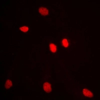
ICC/IF analysis of formalin-fixed MCF7 cells using GTX54916 ADAR2 antibody. Red : Primary antibody Blue : DAPI Permeabilization : 0.1% Triton X-100 in TBS for 5-10 minutes
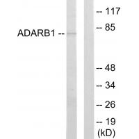
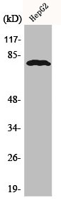
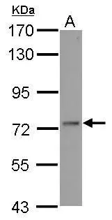
, MCF7 (B), mouse brain (C), rat brain (D) whole cell lysates using GTX54916 ADAR2 antibody.")
")
ICC/IF analysis of formalin-fixed MCF7 cells using GTX54916 ADAR2 antibody. Red : Primary antibody Blue : DAPI Permeabilization : 0.1% Triton X-100 in TBS for 5-10 minutes
ADAR2 antibody
GTX54916
ApplicationsImmunoFluorescence, Western Blot, ImmunoCytoChemistry, ImmunoHistoChemistry, ImmunoHistoChemistry Paraffin
Product group Antibodies
ReactivityHuman, Mouse, Rat
TargetADARB1
Overview
- SupplierGeneTex
- Product NameADAR2 antibody
- Delivery Days Customer9
- Application Supplier NoteWB: 1:500 - 1:1000. ICC/IF: 1:100 - 1:500. IHC-P: 1:100 - 1:200. *Optimal dilutions/concentrations should be determined by the researcher.Not tested in other applications.
- ApplicationsImmunoFluorescence, Western Blot, ImmunoCytoChemistry, ImmunoHistoChemistry, ImmunoHistoChemistry Paraffin
- CertificationResearch Use Only
- ClonalityPolyclonal
- ConjugateUnconjugated
- Gene ID104
- Target nameADARB1
- Target descriptionadenosine deaminase RNA specific B1
- Target synonymsADAR2, DRABA2, DRADA2, NEDHYMS, RED1, double-stranded RNA-specific editase 1, RNA editing deaminase 1, RNA-editing enzyme 1, adenosine deaminase, RNA-specific, B1 (homolog of rat RED1), dsRNA adenosine deaminase DRADA2
- HostRabbit
- IsotypeIgG
- Protein IDP78563
- Protein NameDouble-stranded RNA-specific editase 1
- Scientific DescriptionThis gene encodes the enzyme responsible for pre-mRNA editing of the glutamate receptor subunit B by site-specific deamination of adenosines. Studies in rat found that this enzyme acted on its own pre-mRNA molecules to convert an AA dinucleotide to an AI dinucleotide which resulted in a new splice site. Alternative splicing of this gene results in several transcript variants, some of which have been characterized by the presence or absence of an ALU cassette insert and a short or long C-terminal region. [provided by RefSeq, Jul 2008]
- ReactivityHuman, Mouse, Rat
- Storage Instruction-20°C or -80°C,2°C to 8°C
- UNSPSC41116161
References
- Detection of transcriptome-wide microRNA-target interactions in single cells with agoTRIBE.Read this paper
Datasheet
Related products
Product group Antibodies
Anti-ADARB1 Antibody144-64636
ApplicationsWestern Blot
ReactivityHuman, Mouse, Rat
TargetADARB1
- SizePrice
Product group Antibodies
Anti-ADARB1 AntibodyA42124
ApplicationsWestern Blot
ReactivityHuman, Mouse, Rat
- SizePrice

Product group Antibodies
Anti-RED1/ADARB1 Antibody Picoband(r)A01810-2-CARRIER-FREE
ApplicationsFlow Cytometry, Western Blot, ELISA
ReactivityHuman, Mouse, Rat
TargetADARB1
- SizePrice
Product group Antibodies
ADARB1 AntibodyCSB-PA000821
ApplicationsWestern Blot, ELISA, ImmunoHistoChemistry
ReactivityHuman, Mouse, Rat
TargetADARB1
- SizePrice
Product group Antibodies

ApplicationsWestern Blot, ELISA, ImmunoHistoChemistry
ReactivityHuman, Mouse, Rat
TargetADARB1
- SizePrice
Product group Antibodies
ADAR2 antibodyGTX114237
ApplicationsImmunoFluorescence, Western Blot, ImmunoCytoChemistry
ReactivityHuman
TargetADARB1
- SizePrice

Product group Antibodies
Anti-ADARB1 AntibodyHPA018277
ApplicationsImmunoHistoChemistry
ReactivityHuman
TargetADARB1
- SizePrice